Neves H et al. (MAY 2006)
Stem cells (Dayton,Ohio) 24 5 1328--37
Effects of Delta1 and Jagged1 on early human hematopoiesis: correlation with expression of notch signaling-related genes in CD34+ cells.
It has been shown that Notch signaling mediated by ligands of both Jagged and Delta families expands the hematopoietic stem cell compartment while blocking or delaying terminal myeloid differentiation. Here we show that Delta1- and Jagged1-expressing stromal cells have distinct effects on the clonogenic and differentiation capacities of human CD34(+) CD38(+) cells. Jagged1 increases the number of bipotent colony-forming unit-granulocyte macrophage (CFU-GM) and unipotent progenitors (CFU-granulocytes and CFU-macrophages),without quantitatively affecting terminal cell differentiation,whereas Delta1 reduces the number of CFU-GM and differentiated monocytic cells. Expression analysis of genes coding for Notch receptors,Notch targets,and Notch signaling modulators in supernatant CD34(+) cells arising upon contact with Jagged1 and Delta1 shows dynamic and differential gene expression profiles over time. At early time points,modest upregulation of Notch1,Notch3,and Hes1 was observed in Jagged1-CD34(+) cells,whereas those in contact with Delta1 strikingly upregulated Notch3 and Hes1. Later,myeloid progenitors with strong clonogenic potential emerging upon contact with Jagged1 upregulated Notch1 and Deltex and downregulated Notch signaling modulators,whereas T/NK progenitors originated by Delta1 strikingly upregulated Notch3 and Deltex and,to a lesser extent,Hes1,Lunatic Fringe,and Numb. Together,the data unravel previously unrecognized expression patterns of Notch signaling-related genes in CD34(+) CD38(+) cells as they develop in Jagged1- or Delta1-stromal cell environments,which appear to reflect sequential maturational stages of CD34(+) cells into distinct cell lineages.
View Publication
产品号#:
04435
04445
产品名:
MethoCult™ H4435 Enriched
MethoCult™ H4435 Enriched
Goel A et al. (MAY 2006)
Blood 107 10 4063--70
Synergistic activity of the proteasome inhibitor PS-341 with non-myeloablative 153-Sm-EDTMP skeletally targeted radiotherapy in an orthotopic model of multiple myeloma.
Multiple myeloma is a highly radiosensitive skeletal malignancy,but bone-seeking radionuclides have not yet found their place in disease management. We previously reported that the proteasome inhibitor PS-341 selectively sensitizes myeloma cells to the lethal effects of ionizing radiation. To extend these observations to an in vivo model,we combined PS-341 with the bone-seeking radionuclide 153-Sm-EDTMP. In vitro clonogenic assays demonstrated synergistic killing of myeloma cells exposed to both PS-341 and 153-Sm-EDTMP. Using the orthotopic,syngeneic 5TGM1 myeloma model,the median survivals of mice treated with saline,2 doses of PS-341 (0.5 mg/kg),or a single nonmyeloablative dose of 153-Sm-EDTMP (22.5 MBq) were 21,22,and 28 days,respectively. In contrast,mice treated with combination therapy comprising 2 doses of PS-341 (0.5 mg/kg),1 day prior to and 1 day following 153-Sm-EDTMP (22.5 MBq) showed a significantly prolonged median survival of 49 days (P textless .001). In addition to prolonged survival,this treatment combination yielded reduced clonogenicity of bone marrow-resident 5TGM1 cells,reduced serum myeloma-associated paraprotein levels,and better preservation of bone mineral density. Myelosuppression,determined by peripheral blood cell counts and clonogenicity assays of hematopoietic progenitors,did not differ between animals treated with 153-Sm-EDTMP alone versus those treated with the combination of PS-341 plus 153-Sm-EDTMP. PS-341 is a potent,selective in vivo radiosensitizer that may substantially affect the efficacy of skeletal-targeted radiotherapy in multiple myeloma.
View Publication
产品号#:
04236
产品名:
MethoCult™ SF H4236
Maes C et al. (MAY 2006)
The Journal of clinical investigation 116 5 1230--42
Placental growth factor mediates mesenchymal cell development, cartilage turnover, and bone remodeling during fracture repair.
Current therapies for delayed- or nonunion bone fractures are still largely ineffective. Previous studies indicated that the VEGF homolog placental growth factor (PlGF) has a more significant role in disease than in health. Therefore we investigated the role of PlGF in a model of semi-stabilized bone fracture healing. Fracture repair in mice lacking PlGF was impaired and characterized by a massive accumulation of cartilage in the callus,reminiscent of delayed- or nonunion fractures. PlGF was required for the early recruitment of inflammatory cells and the vascularization of the fracture wound. Interestingly,however,PlGF also played a role in the subsequent stages of the repair process. Indeed in vivo and in vitro findings indicated that PlGF induced the proliferation and osteogenic differentiation of mesenchymal progenitors and stimulated cartilage turnover by particular MMPs. Later in the process,PlGF was required for the remodeling of the newly formed bone by stimulating osteoclast differentiation. As PlGF expression was increased throughout the process of bone repair and all the important cell types involved expressed its receptor VEGFR-1,the present data suggest that PlGF is required for mediating and coordinating the key aspects of fracture repair. Therefore PlGF may potentially offer therapeutic advantages for fracture repair.
View Publication
产品号#:
03534
03334
03434
03444
18753
18753RF
产品名:
MethoCult™ GF M3534
MethoCult™ M3334
MethoCult™ GF M3434
MethoCult™ GF M3434
Baba Y et al. (AUG 2006)
Journal of immunology (Baltimore,Md. : 1950) 177 4 2294--303
Constitutively active beta-catenin promotes expansion of multipotent hematopoietic progenitors in culture.
This study was designed to investigate one component of the Wnt/beta-catenin signaling pathway that has been implicated in stem cell self-renewal. Retroviral-mediated introduction of stable beta-catenin to primitive murine bone marrow cells allowed the expansion of multipotential c-Kit(low)Sca-1(low/-)CD19(-) CD11b/Mac-1(-)Flk-2(-)CD43(+)AA4.1(+)NK1.1(-)CD3(-)CD11c(-)Gr-1(-)CD45R/B220(+) cells in the presence of stromal cells and cytokines. They generated myeloid,T,and B lineage lymphoid cells in culture,but had no T lymphopoietic potential when transplanted. Stem cell factor and IL-6 were found to be minimal requirements for long-term,stromal-free propagation,and a beta-catenin-transduced cell line was maintained for 5 mo with these defined conditions. Although multipotential and responsive to many normal stimuli in culture,it was unable to engraft several types of irradiated recipients. These findings support previous studies that have implicated the canonical Wnt pathway signaling in regulation of multipotent progenitors. In addition,we demonstrate how it may be experimentally manipulated to generate valuable cell lines.
View Publication
产品号#:
03434
03444
产品名:
MethoCult™ GF M3434
MethoCult™ GF M3434
Feng R et al. (MAR 2007)
Blood 109 5 2130--8
SDX-308, a nonsteroidal anti-inflammatory agent, inhibits NF-kappaB activity, resulting in strong inhibition of osteoclast formation/activity and multiple myeloma cell growth.
Multiple myeloma is characterized by increased osteoclast activity that results in bone destruction and lytic lesions. With the prolonged overall patient survival achieved by new treatment modalities,additional drugs are required to inhibit bone destruction. We focused on a novel and more potent structural analog of the nonsteroidal anti-inflammatory drug etodolac,known as SDX-308,and its effects on osteoclastogenesis and multiple myeloma cells. SDX-101 is another structural analog of etodolac that is already used in clinical trials for the treatment of B-cell chronic lymphocytic leukemia (B-CLL). Compared with SDX-101,a 10-fold lower concentration of SDX-308 induced potent (60%-80%) inhibition of osteoclast formation,and a 10- to 100-fold lower concentration inhibited multiple myeloma cell proliferation. Bone resorption was completely inhibited by SDX-308,as determined in dentin-based bone resorption assays. SDX-308 decreased constitutive and RANKL-stimulated NF-kappaB activation and osteoclast formation in an osteoclast cellular model,RAW 264.7. SDX-308 effectively suppressed TNF-alpha-induced IKK-gamma and IkappaB-alpha phosphorylation and degradation and subsequent NF-kappaB activation in human multiple myeloma cells. These results indicate that SDX-308 effectively inhibits multiple myeloma cell proliferation and osteoclast activity,potentially by controlling NF-kappaB activation signaling. We propose that SDX-308 is a promising therapeutic candidate to inhibit multiple myeloma growth and osteoclast activity and that it should receive attention for further study.
View Publication
产品号#:
04434
04444
产品名:
MethoCult™ H4434 Classic
MethoCult™ H4434 Classic
Bruserud &O et al. (MAR 2007)
Haematologica 92 3 332--41
Subclassification of patients with acute myelogenous leukemia based on chemokine responsiveness and constitutive chemokine release by their leukemic cells.
BACKGROUND AND OBJECTIVES: Chemokines are soluble mediators involved in angiogenesis,cellular growth control and immunomodulation. In the present study we investigated the effects of various chemokines on proliferation of acute myelogenous leukemia (AML) cells and constitutive chemokine release by primary AML cells. DESIGN AND METHODS: Native human AML cells derived from 68 consecutive patients were cultured in vitro. We investigated AML cell proliferation (3H-thymidine incorporation,colony formation),chemokine receptor expression,constitutive chemokine release and chemotaxis of normal peripheral blood mononuclear cells. RESULTS: Exogenous chemokines usually did not have any effect on AML blast proliferation in the absence of hematopoietic growth factors,but when investigating growth factor-dependent (interleukin 3 + granulocyte-macrophage colony-stimulating factor + stem cell factor) proliferation in suspension cultures the following patient subsets were identified: (i) patients whose cells showed chemokine-induced growth enhancement (8 patients); (ii) divergent effects on proliferation (15 patients); and (iii) no effect (most patients). These patient subsets did not differ in chemokine receptor expression,but,compared to CD34- AML cells,CD34+ cells showed higher expression of several receptors. Chemokines also increased the proliferation of clonogenic AML cells from the first subset of patients. Furthermore,a broad constitutive chemokine release profile was detected for most patients,and the following chemokine clusters could be identified: CCL2-4/CXCL1/8,CCL5/CXCL9-11 (possibly also CCL23) and CCL13/17/22/24/CXCL5 (possibly also CXCL6). Only the CCL2-4/CXCL1/8 cluster showed significant correlations between corresponding mRNA levels and NFkB levels/activation. The chemotaxis of normal immunocompetent cells for patients without constitutive chemokine release was observed to be decreased. INTERPRETATION AND CONCLUSIONS: Differences in chemokine responsiveness as well as chemokine release contribute to patient heterogeneity in AML. Patients with AML can be classified into distinct subsets according to their chemokine responsiveness and chemokine release profile.
View Publication

 EasySep™小鼠TIL(CD45)正选试剂盒


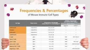 挂图Frequencies and Percentages of Mouse Immune Cell Types List of the frequencies of over 25 immune cell types in C57BL/6 mice

 沪公网安备31010102008431号