Neutrophil survival and c-kit(+)-progenitor proliferation in Staphylococcus aureus-infected skin wounds promote resolution.
Polymorphonuclear neutrophils (PMNs) are critical for the formation,maintenance,and resolution of bacterial abscesses. However,the mechanisms that regulate PMN survival and proliferation during the evolution of an abscess are not well defined. Using a mouse model of Staphylococcus aureus abscess formation within a cutaneous wound,combined with real-time imaging of genetically tagged PMNs,we observed that a high bacterial burden elicited a sustained mobilization of PMNs from the bone marrow to the infected wound,where their lifespan was markedly extended. A continuous rise in wound PMN number,which was not accounted for by trafficking from the bone marrow or by prolonged survival,was correlated with the homing of c-kit(+)-progenitor cells from the blood to the wound,where they proliferated and formed mature PMNs. Furthermore,by blocking their recruitment with an antibody to c-kit,which severely limited the proliferation of mature PMNs in the wound and shortened mouse survival,we confirmed that progenitor cells are not only important contributors to PMN expansion in the wound,but are also functionally important for immune protection. We conclude that the abscess environment provides a niche capable of regulating PMN survival and local proliferation of bone marrow-derived c-kit(+)-progenitor cells.
View Publication
产品号#:
03434
03444
产品名:
MethoCult™ GF M3434
MethoCult™ GF M3434
Yoshimi A et al. (MAR 2011)
Blood 117 13 3617--28
Evi1 represses PTEN expression and activates PI3K/AKT/mTOR via interactions with polycomb proteins.
Evi1 (ecotropic viral integration site 1) is essential for proliferation of hematopoietic stem cells and implicated in the development of myeloid disorders. Particularly,high Evi1 expression defines one of the largest clusters in acute myeloid leukemia and is significantly associated with extremely poor prognosis. However,mechanistic basis of Evi1-mediated leukemogenesis has not been fully elucidated. Here,we show that Evi1 directly represses phosphatase and tensin homologue deleted on chromosome 10 (PTEN) transcription in the murine bone marrow,which leads to activation of AKT/mammalian target of rapamycin (mTOR) signaling. In a murine bone marrow transplantation model,Evi1 leukemia showed modestly increased sensitivity to an mTOR inhibitor rapamycin. Furthermore,we found that Evi1 binds to several polycomb group proteins and recruits polycomb repressive complexes for PTEN down-regulation,which shows a novel epigenetic mechanism of AKT/mTOR activation in leukemia. Expression analyses and ChIPassays with human samples indicate that our findings in mice models are recapitulated in human leukemic cells. Dependence of Evi1-expressing leukemic cells on AKT/mTOR signaling provides the first example of targeted therapeutic modalities that suppress the leukemogenic activity of Evi1. The PTEN/AKT/mTOR signaling pathway and the Evi1-polycomb interaction can be promising therapeutic targets for leukemia with activated Evi1.
View Publication
产品号#:
03434
03444
产品名:
MethoCult™ GF M3434
MethoCult™ GF M3434
Zhao Z et al. (JUL 2010)
Genes & development 24 13 1389--402
p53 loss promotes acute myeloid leukemia by enabling aberrant self-renewal.
The p53 tumor suppressor limits proliferation in response to cellular stress through several mechanisms. Here,we test whether the recently described ability of p53 to limit stem cell self-renewal suppresses tumorigenesis in acute myeloid leukemia (AML),an aggressive cancer in which p53 mutations are associated with drug resistance and adverse outcome. Our approach combined mosaic mouse models,Cre-lox technology,and in vivo RNAi to disable p53 and simultaneously activate endogenous Kras(G12D)-a common AML lesion that promotes proliferation but not self-renewal. We show that p53 inactivation strongly cooperates with oncogenic Kras(G12D) to induce aggressive AML,while both lesions on their own induce T-cell malignancies with long latency. This synergy is based on a pivotal role of p53 in limiting aberrant self-renewal of myeloid progenitor cells,such that loss of p53 counters the deleterious effects of oncogenic Kras on these cells and enables them to self-renew indefinitely. Consequently,myeloid progenitor cells expressing oncogenic Kras and lacking p53 become leukemia-initiating cells,resembling cancer stem cells capable of maintaining AML in vivo. Our results establish an efficient new strategy for interrogating oncogene cooperation,and provide strong evidence that the ability of p53 to limit aberrant self-renewal contributes to its tumor suppressor activity.
View Publication
产品号#:
03534
09600
09650
产品名:
MethoCult™ GF M3534
StemSpan™ SFEM
StemSpan™ SFEM
Mahdipour E et al. (JAN 2011)
Blood 117 3 815--26
Hoxa3 promotes the differentiation of hematopoietic progenitor cells into proangiogenic Gr-1+CD11b+ myeloid cells.
Injury induces the recruitment of bone marrow-derived cells (BMDCs) that contribute to the repair and regeneration process. The behavior of BMDCs in injured tissue has a profound effect on repair,but the regulation of BMDC behavior is poorly understood. Aberrant recruitment/retention of these cells in wounds of diabetic patients and animal models is associated with chronic inflammation and impaired healing. BMD Gr-1(+)CD11b(+) cells function as immune suppressor cells and contribute significantly to tumor-induced neovascularization. Here we report that Gr-1(+)CD11b(+) cells also contribute to injury-induced neovascularization,but show altered recruitment/retention kinetics in the diabetic environment. Moreover,diabetic-derived Gr-1(+)CD11b(+) cells fail to stimulate neovascularization in vivo and have aberrant proliferative,chemotaxis,adhesion,and differentiation potential. Previously we demonstrated that gene transfer of HOXA3 to wounds of diabetic mice is taken up by and expressed by recruited BMDCs. This is associated with a suppressed inflammatory response,enhanced neovascularization,and accelerated wound healing. Here we show that sustained expression of Hoxa3 in diabetic-derived BMD Gr-1(+)CD11b(+) cells reverses their diabetic phenotype. These findings demonstrate that manipulation of adult stem/progenitor cells ex vivo could be used as a potential therapy in patients with impaired wound healing.
View Publication
产品号#:
03434
03444
产品名:
MethoCult™ GF M3434
MethoCult™ GF M3434
Karp JE et al. (MAY 2009)
Blood 113 20 4841--52
Active oral regimen for elderly adults with newly diagnosed acute myelogenous leukemia: a preclinical and phase 1 trial of the farnesyltransferase inhibitor tipifarnib (R115777, Zarnestra) combined with etoposide.
The farnesyltransferase inhibitor tipifarnib exhibits modest activity against acute myelogenous leukemia. To build on these results,we examined the effect of combining tipifarnib with other agents. Tipifarnib inhibited signaling downstream of the farnesylated small G protein Rheb and synergistically enhanced etoposide-induced antiproliferative effects in lymphohematopoietic cell lines and acute myelogenous leukemia isolates. We subsequently conducted a phase 1 trial of tipifarnib plus etoposide in adults over 70 years of age who were not candidates for conventional therapy. A total of 84 patients (median age,77 years) received 224 cycles of oral tipifarnib (300-600 mg twice daily for 14 or 21 days) plus oral etoposide (100-200 mg daily on days 1-3 and 8-10). Dose-limiting toxicities occurred with 21-day tipifarnib. Complete remissions were achieved in 16 of 54 (30%) receiving 14-day tipifarnib versus 5 of 30 (17%) receiving 21-day tipifarnib. Complete remissions occurred in 50% of two 14-day tipifarnib cohorts: 3A (tipifarnib 600,etoposide 100) and 8A (tipifarnib 400,etoposide 200). In vivo,tipifarnib plus etoposide decreased ribosomal S6 protein phosphorylation and increased histone H2AX phosphorylation and apoptosis. Tipifarnib plus etoposide is a promising orally bioavailable regimen that warrants further evaluation in elderly adults who are not candidates for conventional induction chemotherapy. These clinical studies are registered at www.clinicaltrials.gov as NCT00112853.
View Publication

 EasySep™小鼠TIL(CD45)正选试剂盒


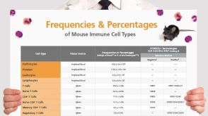 挂图Frequencies and Percentages of Mouse Immune Cell Types List of the frequencies of over 25 immune cell types in C57BL/6 mice


 沪公网安备31010102008431号