Nicolini FE et al. (AUG 2002)
Blood 100 4 1257--64
Expression of a human beta-globin transgene in erythroid cells derived from retrovirally transduced transplantable human fetal liver and cord blood cells.
Transfer of therapeutic genes to human hematopoietic stem cells (HSCs) using complex vectors at clinically relevant efficiencies remains a major challenge. Recently we described a stable retroviral vector that sustains long-term expression of green fluorescent protein (GFP) and a human beta-globin gene in the erythroid progeny of transduced murine HSCs. We now report the efficient transduction of primitive human CD34(+) fetal liver or cord blood cells with this vector and expression of the beta-globin transgene in the erythroid progeny of these human cells for at least 2 months. After growth factor prestimulation and then a 2- to 3-day exposure to the virus,35% to 55% GFP(+) progeny were seen in assays of transduced colony-forming cells,primitive erythroid precursors that generate large numbers of glycophorin A(+) cells in 3-week suspension cultures,and 6-week long-term culture-initiating cells. In immunodeficient mice injected with unselected infected cells,5% to 15% of the human cells regenerated in the marrow (including the erythroid cells) were GFP(+) 3 and 6 weeks after transplantation. Importantly,the numbers of GFP(+) human lymphoid and either granulopoietic or erythroid cells in individual mice 6 weeks after transplantation were significantly correlated,indicative of the initial transduction of human multipotent cells with in vivo repopulating activity. Expression of the transduced beta-globin gene in human cells obtained directly from the mice or after their differentiation into erythroid cells in vitro was demonstrated by reverse transcriptase-polymerase chain reaction using specific primers. These experiments represent a significant step toward the realization of a gene therapy approach for human beta-globin gene disorders.
View Publication
产品号#:
04330
产品名:
MethoCult™ H4330
Sandrin V et al. (AUG 2002)
Blood 100 3 823--32
Lentiviral vectors pseudotyped with a modified RD114 envelope glycoprotein show increased stability in sera and augmented transduction of primary lymphocytes and CD34+ cells derived from human and nonhuman primates.
Generating lentiviral vectors pseudotyped with different viral glycoproteins (GPs) may modulate the physicochemical properties of the vectors,their interaction with the host immune system,and their host range. We have investigated the capacity of a panel of GPs of both retroviral (amphotropic murine leukemia virus [MLV-A]; gibbon ape leukemia virus [GALV]; RD114,feline endogenous virus) and nonretroviral (fowl plague virus [FPV]; Ebola virus [EboV]; vesicular stomatitis virus [VSV]; lymphocytic choriomeningitis virus [LCMV]) origins to pseudotype lentiviral vectors derived from simian immunodeficiency virus (SIVmac251). SIV vectors were efficiently pseudotyped with the FPV hemagglutinin,VSV-G,LCMV,and MLV-A GPs. In contrast,the GALV and RD114 GPs conferred much lower infectivity to the vectors. Capitalizing on the conservation of some structural features in the transmembrane domains and cytoplasmic tails of the incorporation-competent MLV-A GP and in RD114 and GALV GPs,we generated chimeric GPs encoding the extracellular and transmembrane domains of GALV or RD114 GPs fused to the cytoplasmic tail (designated TR) of MLV-A GP. Importantly,SIV-derived vectors pseudotyped with these GALV/TR and RD114/TR GP chimeras had significantly higher titers than vectors coated with the parental GPs. Additionally,RD114/TR-pseudotyped vectors were efficiently concentrated and were resistant to inactivation induced by the complement of both human and macaque sera,indicating that modified RD114 GP-pseudotyped lentiviral vectors may be of particular interest for in vivo gene transfer applications. Furthermore,as compared to vectors pseudotyped with other retroviral GPs or with VSV-G,RD114/TR-pseudotyped vectors showed augmented transduction of human and macaque primary blood lymphocytes and CD34+ cells.
View Publication
产品号#:
02690
09600
09650
产品名:
StemSpan™ CC100
StemSpan™ SFEM
StemSpan™ SFEM
Chen X et al. (SEP 2015)
Stem Cell Research 15 2 395--402
OP9-Lhx2 stromal cells facilitate derivation of hematopoietic progenitors both in vitro and in vivo
Generating engraftable hematopoietic stem cells (HSCs) from pluripotent stem cells (PSCs) is an ideal approach for obtaining induced HSCs for cell therapy. However,the path from PSCs to robustly induced HSCs (iHSCs) in vitro remains elusive. We hypothesize that the modification of hematopoietic niche cells by transcription factors facilitates the derivation of induced HSCs from PSCs. The Lhx2 transcription factor is expressed in fetal liver stromal cells but not in fetal blood cells. Knocking out Lhx2 leads to a fetal hematopoietic defect in a cell non-autonomous role. In this study,we demonstrate that the ectopic expression of Lhx2 in OP9 cells (OP9-Lhx2) accelerates the hematopoietic differentiation of PSCs. OP9-Lhx2 significantly increased the yields of hematopoietic progenitor cells via co-culture with PSCs in vitro. Interestingly,the co-injection of OP9-Lhx2 and PSCs into immune deficient mice also increased the proportion of hematopoietic progenitors via the formation of teratomas. The transplantation of phenotypic HSCs from OP9-Lhx2 teratomas but not from the OP9 control supported a transient repopulating capability. The upregulation of Apln gene by Lhx2 is correlated to the hematopoietic commitment property of OP9-Lhx2. Furthermore,the enforced expression of Apln in OP9 cells significantly increased the hematopoietic differentiation of PSCs. These results indicate that OP9-Lhx2 is a good cell line for regeneration of hematopoietic progenitors both in vitro and in vivo.
View Publication
产品号#:
05850
05857
05870
05875
85850
85857
85870
85875
产品名:
mTeSR™1
mTeSR™1
Lee H-Y et al. (JUN 2015)
Nature 522 7557 474--7
PPAR-α and glucocorticoid receptor synergize to promote erythroid progenitor self-renewal.
Many acute and chronic anaemias,including haemolysis,sepsis and genetic bone marrow failure diseases such as Diamond-Blackfan anaemia,are not treatable with erythropoietin (Epo),because the colony-forming unit erythroid progenitors (CFU-Es) that respond to Epo are either too few in number or are not sensitive enough to Epo to maintain sufficient red blood cell production. Treatment of these anaemias requires a drug that acts at an earlier stage of red cell formation and enhances the formation of Epo-sensitive CFU-E progenitors. Recently,we showed that glucocorticoids specifically stimulate self-renewal of an early erythroid progenitor,burst-forming unit erythroid (BFU-E),and increase the production of terminally differentiated erythroid cells. Here we show that activation of the peroxisome proliferator-activated receptor α (PPAR-α) by the PPAR-α agonists GW7647 and fenofibrate synergizes with the glucocorticoid receptor (GR) to promote BFU-E self-renewal. Over time these agonists greatly increase production of mature red blood cells in cultures of both mouse fetal liver BFU-Es and mobilized human adult CD34(+) peripheral blood progenitors,with a new and effective culture system being used for the human cells that generates normal enucleated reticulocytes. Although Ppara(-/-) mice show no haematological difference from wild-type mice in both normal and phenylhydrazine (PHZ)-induced stress erythropoiesis,PPAR-α agonists facilitate recovery of wild-type but not Ppara(-/-) mice from PHZ-induced acute haemolytic anaemia. We also show that PPAR-α alleviates anaemia in a mouse model of chronic anaemia. Finally,both in control and corticosteroid-treated BFU-E cells,PPAR-α co-occupies many chromatin sites with GR; when activated by PPAR-α agonists,additional PPAR-α is recruited to GR-adjacent sites and presumably facilitates GR-dependent BFU-E self-renewal. Our discovery of the role of PPAR-α agonists in stimulating self-renewal of early erythroid progenitor cells suggests that the clinically tested PPAR-α agonists we used may improve the efficacy of corticosteroids in treating Epo-resistant anaemias.
View Publication
产品号#:
02697
06902
06952
09300
09600
09650
产品名:
StemSpan™ CC110
含有10% 牛血清白蛋白(BSA)的 Iscove's MDM
StemSpan™ SFEM
StemSpan™ SFEM
Kolodziej S et al. (MAY 2014)
Nature communications 5 3995
PADI4 acts as a coactivator of Tal1 by counteracting repressive histone arginine methylation.
The transcription factor Tal1 is a critical activator or repressor of gene expression in hematopoiesis and leukaemia. The mechanism by which Tal1 differentially influences transcription of distinct genes is not fully understood. Here we show that Tal1 interacts with the peptidylarginine deiminase IV (PADI4). We demonstrate that PADI4 can act as an epigenetic coactivator through influencing H3R2me2a. At the Tal1/PADI4 target gene IL6ST the repressive H3R2me2a mark triggered by PRMT6 is counteracted by PADI4,which augments the active H3K4me3 mark and thus increases IL6ST expression. In contrast,at the CTCF promoter PADI4 acts as a repressor. We propose that the influence of PADI4 on IL6ST transcription plays a role in the control of IL6ST expression during lineage differentiation of hematopoietic stem/progenitor cells. These results open the possibility to pharmacologically influence Tal1 in leukaemia.
View Publication
产品号#:
07930
07931
07940
07955
07956
07959
07954
100-1061
07952
产品名:
CryoStor® CS10
CryoStor® CS10
CryoStor® CS10
CryoStor® CS10
CryoStor® CS10
CryoStor® CS10
CryoStor® CS10
Pei S et al. (NOV 2013)
The Journal of biological chemistry 288 47 33542--58
Targeting aberrant glutathione metabolism to eradicate human acute myelogenous leukemia cells.
The development of strategies to eradicate primary human acute myelogenous leukemia (AML) cells is a major challenge to the leukemia research field. In particular,primitive leukemia cells,often termed leukemia stem cells,are typically refractory to many forms of therapy. To investigate improved strategies for targeting of human AML cells we compared the molecular mechanisms regulating oxidative state in primitive (CD34(+)) leukemic versus normal specimens. Our data indicate that CD34(+) AML cells have elevated expression of multiple glutathione pathway regulatory proteins,presumably as a mechanism to compensate for increased oxidative stress in leukemic cells. Consistent with this observation,CD34(+) AML cells have lower levels of reduced glutathione and increased levels of oxidized glutathione compared with normal CD34(+) cells. These findings led us to hypothesize that AML cells will be hypersensitive to inhibition of glutathione metabolism. To test this premise,we identified compounds such as parthenolide (PTL) or piperlongumine that induce almost complete glutathione depletion and severe cell death in CD34(+) AML cells. Importantly,these compounds only induce limited and transient glutathione depletion as well as significantly less toxicity in normal CD34(+) cells. We further determined that PTL perturbs glutathione homeostasis by a multifactorial mechanism,which includes inhibiting key glutathione metabolic enzymes (GCLC and GPX1),as well as direct depletion of glutathione. These findings demonstrate that primitive leukemia cells are uniquely sensitive to agents that target aberrant glutathione metabolism,an intrinsic property of primary human AML cells.
View Publication
产品号#:
07930
07931
07940
07955
07956
07959
07954
100-1061
07952
产品名:
CryoStor® CS10
CryoStor® CS10
CryoStor® CS10
CryoStor® CS10
CryoStor® CS10
CryoStor® CS10
CryoStor® CS10
Iversen PO et al. (JAN 2002)
American journal of physiology. Regulatory,integrative and comparative physiology 282 1 R166--72
Decreased hematopoiesis in bone marrow of mice with congestive heart failure.
Patients with heart failure are predisposed to infections and anemia,possibly due to reduced hematopoiesis. The proinflammatory cytokine tumor necrosis factor-alpha (TNF-alpha) is increased in heart failure,and it inhibits normal hematopoiesis,partly due to apoptosis through the effector molecule Fas. We examined bone marrow progenitor cells of mice with heart failure induced by acute myocardial infarction. The fraction of progenitor cells in mice with heart failure was only approximately 40% of control. Measured with in vitro clonal assays,the proliferative capacity of the progenitor cells in mice with heart failure was reduced to approximately 50% of control. Flow cytometry with specific markers revealed a threefold increase in apoptosis among progenitor cells from mice with heart failure. In these mice,TNF-alpha/Fas expression was increased in bone marrow natural killer (NK) and T cells,and these lymphocytes showed increased cytolytic activity in vitro against progenitor cells. We conclude that the TNF-alpha/Fas pathway in lymphocytes is activated in the bone marrow during heart failure,which may play a pathogenic role in the observed decrease in hematopoiesis.
View Publication
产品号#:
05350
产品名:
Coata G et al. (JAN 2001)
Stem cells (Dayton,Ohio) 19 6 534--42
Prenatal diagnosis of genetic abnormalities using fetal CD34+ stem cells in maternal circulation and evidence they do not affect diagnosis in later pregnancies.
In the present study,we report a new method for enrichment and analysis of fetal CD34+ stem cells after culture in order to determine whether it is feasible for noninvasive prenatal diagnosis. We also determined whether fetal CD34+ stem cells persist in maternal blood after delivery and assessed whether they have an impact on noninvasive prenatal diagnosis of genetic abnormalities. Peripheral blood samples were obtained from 35 pregnant women,13 non-pregnant women who had given birth to male offsprings,12 women who had never been pregnant,and eight pregnant women with male fetuses. CD34+ stem cells were enriched and either cultured for prenatal diagnosis or analyzed with fluorescence in situ hybridization (FISH)/polymerase chain reaction (PCR) to determine peristance in maternal blood. Fetal/maternal cells can be isolated and grown in vitro" to provide enough cells for a more accurate fetal sex or aneuploid prediction than is provided by unenriched and uncultured CD34+ stem cells. The presence of fetal cells in maternal blood samples from mothers who had given birth to male offspring was found in 3 of 13 blood samples. PCR was positive for Y chromosome in one woman who had never been pregnant. Analysis of cultured CD34+ stem cells from mothers with Y PCR positivity did not detect any male cells in any samples. Even if PCR positivity is due to persistence of fetal stem cells from previous pregnancies�
View Publication

 EasySep™小鼠TIL(CD45)正选试剂盒


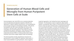 技术公告Generation of Human Blood Cells and Microglia from Human Pluripotent Stem Cells at Scale
 实验方案Optimizing Delivery Efficiency with Fluorescent Dextran Using the CellPore™ Transfection System

 沪公网安备31010102008431号