Haniffa M et al. (FEB 2009)
The Journal of experimental medicine 206 2 371--85
Differential rates of replacement of human dermal dendritic cells and macrophages during hematopoietic stem cell transplantation.
Animal models of hematopoietic stem cell transplantation have been used to analyze the turnover of bone marrow-derived cells and to demonstrate the critical role of recipient antigen-presenting cells (APC) in graft versus host disease (GVHD). In humans,the phenotype and lineage relationships of myeloid-derived tissue APC remain incompletely understood. It has also been proposed that the risk of acute GVHD,which extends over many months,is related to the protracted survival of certain recipient APC. Human dermis contains three principal subsets of CD45(+)HLA-DR(+) cells: CD1a(+)CD14(-) DC,CD1a(-)CD14(+) DC,and CD1a(-)CD14(+)FXIIIa(+) macrophages. In vitro,each subset has characteristic properties. After transplantation,both CD1a(+) and CD14(+) DC are rapidly depleted and replaced by donor cells,but recipient macrophages can be found in GVHD lesions and may persist for many months. Macrophages isolated from normal dermis secrete proinflammatory cytokines. Although they stimulate little proliferation of naive or memory CD4(+) T cells,macrophages induce cytokine expression in memory CD4(+) T cells and activation and proliferation of CD8(+) T cells. These observations suggest that dermal macrophages and DC are from distinct lineages and that persistent recipient macrophages,although unlikely to initiate alloreactivity,may contribute to GVHD by sustaining the responses of previously activated T cells.
View Publication
Bone Marrow Niches and HSC Fates
A detailed reference on signaling pathways in the bone marrow and how these influence HSC fate decisions; created in partnership with Nature Reviews Immunology and Nature Reviews Molecular Cell Biology
Courteney Lai
Life as a Ph.D. Student with a Passion for Myeloid Leukemia
研究方向:
干细胞生物学
Donahue RE et al. (JAN 2000)
Blood 95 2 445--52
High levels of lymphoid expression of enhanced green fluorescent protein in nonhuman primates transplanted with cytokine-mobilized peripheral blood CD34(+) cells.
We have used a murine retrovirus vector containing an enhanced green fluorescent protein complimentary DNA (EGFP cDNA) to dynamically follow vector-expressing cells in the peripheral blood (PB) of transplanted rhesus macaques. Cytokine mobilized CD34(+) cells were transduced with an amphotropic vector that expressed EGFP and a dihydrofolate reductase cDNA under control of the murine stem cell virus promoter. The transduction protocol used the CH-296 recombinant human fibronectin fragment and relatively high concentrations of the flt-3 ligand and stem cell factor. Following transplantation of the transduced cells,up to 55% EGFP-expressing granulocytes were obtained in the peripheral circulation during the early posttransplant period. This level of myeloid marking,however,decreased to 0.1% or lower within 2 weeks. In contrast,EGFP expression in PB lymphocytes rose from 2%-5% shortly following transplantation to 10% or greater by week 5. After 10 weeks,the level of expression in PB lymphocytes continued to remain at 3%-5% as measured by both flow cytometry and Southern blot analysis,and EGFP expression was observed in CD4(+),CD8(+),CD20(+),and CD16/56(+) lymphocyte subsets. EGFP expression was only transiently detected in red blood cells and platelets soon after transplantation. Such sustained levels of lymphocyte marking may be therapeutic in a number of human gene therapy applications that require targeting of the lymphoid compartment. The transient appearance of EGFP(+) myeloid cells suggests that transduction of a lineage-restricted myeloid progenitor capable of short-term engraftment was obtained with this protocol. (Blood. 2000;95:445-452)
View Publication
产品号#:
04436
04064
04100
04230
04236
04431
04434
04444
04464
04531
04535
04545
04536
04564
04035
04330
04034
04044
04435
04445
04534
04544
产品名:
MethoCult™ SF H4436
MethoCult™ H4034 Optimum 入门试剂盒
MethoCult™ H4100
MethoCult™ H4230
MethoCult™ SF H4236
MethoCult™ H4431
MethoCult™ H4434 Classic
MethoCult™ H4434 Classic
MethoCult™ H4434 Classic 套装
MethoCult™ H4531
MethoCult™ H4535 Enriched,不含EPO
MethoCult™ H4535 Enriched,不含EPO
MethoCult™ SF H4536
MethoCult™ H4534 Classic 无 EPO 入门试剂盒
MethoCult™ 不含EPO的H4035 Optimum
MethoCult™ H4330
MethoCult™ H4034 Optimum
MethoCult™ H4034 Optimum
MethoCult™ H4435 Enriched
MethoCult™ H4435 Enriched
MethoCult™ H4534 Classic(不含 EPO)
MethoCult™ H4534 Classic(不含 EPO)
Dobo I et al. (DEC 1999)
Journal of hematotherapy & stem cell research 8 6 601--7
Endogenous erythroid and megakaryocytic colony formation in serum-free, cytokine-free collagen gels.
We studied the suitability of collagen-based semisolid medium for assay of endogenous erythroid colony formation performed in myeloproliferative disorders. Bone marrow (BM) mononuclear cells (MNC) from 103 patients suspected of having polycythemia vera (PV,76 patients) or essential thrombocythemia (ET,27 patients) were grown in collagen-based,serum-free,cytokine-free semisolid medium. Colony analysis at day 8 or 10 showed that this collagen assay is specific,as endogenous growth of erythroid colonies was never observed in cultures of 16 healthy donors and 6 chronic myelogenous leukemia (CML) patients. Endogenous erythroid colony formation was observed in 53.3% of patients suspected of PV,with only 15.4% of positive cultures for patients with 1 minor PV criterion and 72% (p = 0.009) of positive cultures for patients with textgreater or =2 minor or 1 major PV criterion. Similarly,endogenous growth of erythroid colonies was found in 44.4% of patients suspected of ET,with 31.6% of positive cultures for patients with 1 ET criterion versus 75% for patients with textgreater or =2 ET criteria. In addition,we found that in collagen gels,tests of erythropoietin (EPO) hypersensitivity in the presence of 0.01 or 0.05 U/ml of EPO and tests of endogenous colony-forming units-megakaryocyte (CFU-MK) formation cannot be used to detect PV or ET,as these tests were positive for,respectively,21.4% and 50% of healthy donors and 83% and 50% of CML patients. A retrospective analysis suggests that collagen assays are more sensitive than methylcellulose assays to assess endogenous growth of erythroid colonies. In summary,serum-free collagen-based colony assays are simple and reliable assays of endogenous growth of erythroid colonies in myeloproliferative diseases. They also appear to be more sensitive than methylcellulose-based assays.
View Publication

 EasySep™小鼠TIL(CD45)正选试剂盒


 技术窍门人造血干细胞和祖细胞表型的鉴定
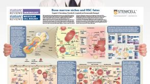 挂图Bone Marrow Niches and HSC Fates A detailed reference on signaling pathways in the bone marrow and how these influence HSC fate decisions; created in partnership with Nature Reviews Immunology and Nature Reviews Molecular Cell Biology


 沪公网安备31010102008431号