Kandilci A and Grosveld GC (AUG 2009)
Blood 114 8 1596--606
Reintroduction of CEBPA in MN1-overexpressing hematopoietic cells prevents their hyperproliferation and restores myeloid differentiation.
Forced expression of MN1 in primitive mouse hematopoietic cells causes acute myeloid leukemia and impairs all-trans retinoic acid-induced granulocytic differentiation. Here,we studied the effects of MN1 on myeloid differentiation and proliferation using primary human CD34(+) hematopoietic cells,lineage-depleted mouse bone marrow cells,and bipotential (granulocytic/monocytic) human acute myeloid leukemia cell lines. We show that exogenous MN1 stimulated the growth of CD34(+) cells,which was accompanied by enhanced survival and increased cell cycle traverse in cultures supporting progenitor cell growth. Forced MN1 expression impaired both granulocytic and monocytic differentiation in vitro in primary hematopoietic cells and acute myeloid leukemia cell lines. Endogenous MN1 expression was higher in human CD34(+) cells compared with both primary and in vitro-differentiated monocytes and granulocytes. Microarray and real-time reverse-transcribed polymerase chain reaction analysis of MN1-overexpressing CD34(+) cells showed down-regulation of CEBPA and its downstream target genes. Reintroduction of conditional and constitutive CEBPA overcame the effects of MN1 on myeloid differentiation and inhibited MN1-induced proliferation in vitro. These results indicate that down-regulation of CEBPA activity contributes to MN1-modulated proliferation and impaired myeloid differentiation of hematopoietic cells.
View Publication
产品号#:
70002
70002.1
70002.2
70002.3
70002.4
70002.5
产品名:
Bueno C et al. (SEP 2009)
Carcinogenesis 30 9 1628--37
Etoposide induces MLL rearrangements and other chromosomal abnormalities in human embryonic stem cells.
MLL rearrangements are hallmark genetic abnormalities in infant leukemia known to arise in utero. They can be induced during human prenatal development upon exposure to etoposide. We also hypothesize that chronic exposure to etoposide might render cells more susceptible to other genomic insults. Here,for the first time,human embryonic stem cells (hESCs) were used as a model to test the effects of etoposide on human early embryonic development. We addressed whether: (i) low doses of etoposide promote MLL rearrangements in hESCs and hESCs-derived hematopoietic cells; (ii) MLL rearrangements are sufficient to confer hESCs with a selective growth advantage and (iii) continuous exposure to low doses of etoposide induces hESCs to acquire other chromosomal abnormalities. In contrast to cord blood-derived CD34(+) and hESC-derived hematopoietic cells,exposure of undifferentiated hESCs to a single low dose of etoposide induced a pronounced cell death. Etoposide induced MLL rearrangements in hESCs and their hematopoietic derivatives. After long-term culture,the proportion of hESCs harboring MLL rearrangements diminished and neither cell cycle variations nor genomic abnormalities were observed in the etoposide-treated hESCs,suggesting that MLL rearrangements are insufficient to confer hESCs with a selective proliferation/survival advantage. However,continuous exposure to etoposide induced MLL breaks and primed hESCs to acquire other major karyotypic abnormalities. These data show that chronic exposure of developmentally early stem cells to etoposide induces MLL rearrangements and make hESCs more prone to acquire other chromosomal abnormalities than postnatal CD34(+) cells,linking embryonic genotoxic exposure to genomic instability.
View Publication
产品号#:
07800
07850
09600
09650
84434
84444
产品名:
氯化铵溶液
氯化铵溶液
StemSpan™ SFEM
StemSpan™ SFEM
Uchida N et al. (OCT 2009)
Journal of virology 83 19 9854--62
Development of a human immunodeficiency virus type 1-based lentiviral vector that allows efficient transduction of both human and rhesus blood cells.
Human immunodeficiency virus type 1 (HIV-1) vectors transduce rhesus blood cells poorly due to a species-specific block by TRIM5alpha and APOBEC3G,which target HIV-1 capsid and viral infectivity factor (Vif),respectively. We sought to develop a lentiviral vector capable of transducing both human and rhesus blood cells by combining components of both HIV-1 and simian immunodeficiency virus (SIV),including SIV capsid (sCA) and SIV Vif. A chimeric HIV-1 vector including sCA (chiHIV) was superior to the conventional SIV in transducing a human blood cell line and superior to the conventional HIV-1 vector in transducing a rhesus blood cell line. Among human CD34(+) hematopoietic stem cells (HSCs),the chiHIV and HIV-1 vectors showed similar transduction efficiencies; in rhesus CD34(+) HSCs,the chiHIV vector yielded superior transduction rates. In in vivo competitive repopulation experiments with two rhesus macaques,the chiHIV vector demonstrated superior marking levels over the conventional HIV-1 vector in all blood lineages (first rhesus,15 to 30% versus 1 to 5%; second rhesus,7 to 15% versus 0.5 to 2%,respectively) 3 to 7 months postinfusion. In summary,we have developed an HIV-1-based lentiviral vector system that should allow comprehensive preclinical testing of HIV-1-based therapeutic vectors in the rhesus macaque model with eventual clinical application.
View Publication
产品号#:
04230
60132
产品名:
MethoCult™ H4230
抗恒河猴红细胞抗体,clone T3G6
Sauer AV et al. (OCT 2009)
Blood 114 15 3216--26
ADA-deficient SCID is associated with a specific microenvironment and bone phenotype characterized by RANKL/OPG imbalance and osteoblast insufficiency.
Adenosine deaminase (ADA) deficiency is a disorder of the purine metabolism leading to combined immunodeficiency and systemic alterations,including skeletal abnormalities. We report that ADA deficiency in mice causes a specific bone phenotype characterized by alterations of structural properties and impaired mechanical competence. These alterations are the combined result of an imbalanced receptor activator of nuclear factor-kappaB ligand (RANKL)/osteoprotegerin axis,causing decreased osteoclastogenesis and an intrinsic defect of osteoblast function with subsequent low bone formation. In vitro,osteoblasts lacking ADA displayed an altered transcriptional profile and growth reduction. Furthermore,the bone marrow microenvironment of ADA-deficient mice showed a reduced capacity to support in vitro and in vivo hematopoiesis. Treatment of ADA-deficient neonatal mice with enzyme replacement therapy,bone marrow transplantation,or gene therapy resulted in full recovery of the altered bone parameters. Remarkably,untreated ADA-severe combined immunodeficiency patients showed a similar imbalance in RANKL/osteoprotegerin levels alongside severe growth retardation. Gene therapy with ADA-transduced hematopoietic stem cells increased serum RANKL levels and children's growth. Our results indicate that the ADA metabolism represents a crucial modulatory factor of bone cell activities and remodeling.
View Publication

 EasySep™小鼠TIL(CD45)正选试剂盒


 实验方案Optimizing Delivery Efficiency with Fluorescent Dextran Using the CellPore™ Transfection System
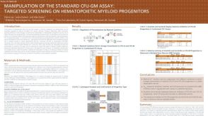 科学海报Manipulation of the Standard CFU-GM Assay Targeted Screening of Hematopoietic Myeloid Progenitors


 沪公网安备31010102008431号