Singbrant S et al. (MAY 2011)
Blood 117 21 5631--42
Erythropoietin couples erythropoiesis, B-lymphopoiesis, and bone homeostasis within the bone marrow microenvironment.
Erythropoietin (Epo) has been used in the treatment of anemia resulting from numerous etiologies,including renal disease and cancer. However,its effects are controversial and the expression pattern of the Epo receptor (Epo-R) is debated. Using in vivo lineage tracing,we document that within the hematopoietic and mesenchymal lineage,expression of Epo-R is essentially restricted to erythroid lineage cells. As expected,adult mice treated with a clinically relevant dose of Epo had expanded erythropoiesis because of amplification of committed erythroid precursors. Surprisingly,we also found that Epo induced a rapid 26% loss of the trabecular bone volume and impaired B-lymphopoiesis within the bone marrow microenvironment. Despite the loss of trabecular bone,hematopoietic stem cell populations were unaffected. Inhibition of the osteoclast activity with bisphosphonate therapy blocked the Epo-induced bone loss. Intriguingly,bisphosphonate treatment also reduced the magnitude of the erythroid response to Epo. These data demonstrate a previously unrecognized in vivo regulatory network coordinating erythropoiesis,B-lymphopoiesis,and skeletal homeostasis. Importantly,these findings may be relevant to the clinical application of Epo.
View Publication
产品号#:
03334
09500
产品名:
MethoCult™ M3334
BIT 9500血清替代物
Martinelli P et al. (JUN 2011)
Blood 117 24 6617--26
The lymphoma-associated NPM-ALK oncogene elicits a p16INK4a/pRb-dependent tumor-suppressive pathway.
Oncogene-induced senescence (OIS) is a barrier for tumor development. Oncogene-dependent DNA damage and activation of the ARF/p53 pathway play a central role in OIS and,accordingly,ARF and p53 are frequently mutated in human cancer. A number of leukemia/lymphoma-initiating oncogenes,however,inhibit ARF/p53 and only infrequently select for ARF or p53 mutations,suggesting the involvement of other tumor-suppressive pathways. We report that NPM-ALK,the initiating oncogene of anaplastic large cell lymphomas (ALCLs),induces DNA damage and irreversibly arrests the cell cycle of primary fibroblasts and hematopoietic progenitors. This effect is associated with inhibition of p53 and is caused by activation of the p16INK4a/pRb tumor-suppressive pathway. Analysis of NPM-ALK lymphomagenesis in transgenic mice showed p16INK4a-dependent accumulation of senescent cells in premalignant lesions and decreased tumor latency in the absence of p16INK4a. Accordingly,human ALCLs showed no expression of either p16INK4a or pRb. Up-regulation of the histone-demethylase Jmjd3 and de-methylation at the p16INK4a promoter contributed to the effect of NPM-ALK on p16INK4a,which was transcriptionally regulated. These data demonstrate that p16INK4a/pRb may function as an alternative pathway of oncogene-induced senescence,and suggest that the reactivation of p16INK4a expression might be a novel strategy to restore the senescence program in some tumors.
View Publication
SALL4 is a robust stimulator for the expansion of hematopoietic stem cells.
HSCs are rare cells that have the unique ability to self-renew and differentiate into cells of all hematopoietic lineages. The lack of donors and current inability to rapidly and efficiently expand HSCs are roadblocks in the development of successful cell therapies. Thus,the challenge of ex vivo human HSC expansion remains a fertile and critically important area of investigation. Here,we show that either SALL4A- or SALL4B-transduced human HSCs obtained from the mobilized peripheral blood are capable of rapid and efficient expansion ex vivo by textgreater10 000-fold for both CD34(+)/CD38(-) and CD34(+)/CD38(+) cells in the presence of appropriate cytokines. We found that these cells retained hematopoietic precursor cell immunophenotypes and morphology as well as normal in vitro or vivo potential for differentiation. The SALL4-mediated expansion was associated with enhanced stem cell engraftment and long-term repopulation capacity in vivo. Also,we demonstrated that constitutive expression of SALL4 inhibited granulocytic differentiation and permitted expansion of undifferentiated cells in 32D myeloid progenitors. Furthermore,a TAT-SALL4B fusion rapidly expanded CD34(+) cells,and it is thus feasible to translate this study into the clinical setting. Our findings provide a new avenue for investigating mechanisms of stem cell self-renewal and achieving clinically significant expansion of human HSCs.
View Publication
产品号#:
09600
09650
产品名:
StemSpan™ SFEM
StemSpan™ SFEM
Lu S-J et al. (JUL 2013)
Regenerative medicine 8 4 413--424
3D microcarrier system for efficient differentiation of human pluripotent stem cells into hematopoietic cells without feeders and serum [corrected].
BACKGROUND Human embryonic stem cells (hESCs) have been derived and maintained on mouse embryonic fibroblast feeders to keep their undifferentiated status. To realize their clinical potential,a feeder-free and scalable system for large scale production of hESCs and their differentiated derivatives is required. MATERIALS & METHODS hESCs were cultured and passaged on serum/feeder-free 3D microcarriers for five passages. For embryoid body (EB) formation and hemangioblast differentiation,the medium for 3D microcarriers was directly switched to EB medium. RESULTS hESCs on 3D microcarriers maintained pluripotency and formed EBs,which were ten-times more efficient than hESCs cultured under 2D feeder-free conditions (0.11 ± 0.03 EB cells/hESC input 2D vs 1.19 ± 0.32 EB cells/hESC input 3D). After replating,EB cells from 3D culture readily developed into hemangioblasts with the potential to differentiate into hematopoietic and endothelial cells. Furthermore,this 3D system can also be adapted to human induced pluripotent stem cells,which generate functional hemangioblasts with high efficiency. CONCLUSION This 3D serum- and stromal-free microcarrier system is important for future clinical applications,with the potential of developing to a GMP-compatible scalable system.
View Publication

 EasySep™小鼠TIL(CD45)正选试剂盒


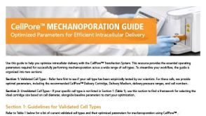
 实验方案Optimizing Delivery Efficiency with Fluorescent Dextran Using the CellPore™ Transfection System


 沪公网安备31010102008431号