Kurita R et al. (SEP 2006)
Stem cells (Dayton,Ohio) 24 9 2014--22
Tal1/Scl gene transduction using a lentiviral vector stimulates highly efficient hematopoietic cell differentiation from common marmoset (Callithrix jacchus) embryonic stem cells.
The development of embryonic stem cell (ESC) therapies requires the establishment of efficient methods to differentiate ESCs into specific cell lineages. Here,we report the in vitro differentiation of common marmoset (CM) (Callithrix jacchus) ESCs into hematopoietic cells after exogenous gene transfer using vesicular stomatitis virus-glycoprotein-pseudotyped lentiviral vectors. We transduced hematopoietic genes,including tal1/scl,gata1,gata2,hoxB4,and lhx2,into CM ESCs. By immunochemical and morphological analyses,we demonstrated that overexpression of tal1/scl,but not the remaining genes,dramatically increased hematopoiesis of CM ESCs,resulting in multiple blood-cell lineages. Furthermore,flow cytometric analysis demonstrated that CD34,a hematopoietic stem/progenitor cell marker,was highly expressed in tal1/scl-overexpressing embryoid body cells. Similar results were obtained from three independent CM ESC lines. These results suggest that transduction of exogenous tal1/scl cDNA into ESCs is a promising method to induce the efficient differentiation of CM ESCs into hematopoietic stem/progenitor cells.
View Publication
Wunderlich M et al. (SEP 2006)
Blood 108 5 1690--7
Human CD34+ cells expressing the inv(16) fusion protein exhibit a myelomonocytic phenotype with greatly enhanced proliferative ability.
The t(16:16) and inv(16) are associated with FAB M4Eo myeloid leukemias and result in fusion of the CBFB gene to the MYH11 gene (encoding smooth muscle myosin heavy chain [SMMHC]). Knockout of CBFbeta causes embryonic lethality due to lack of definitive hematopoiesis. Although knock-in of CBFB-MYH11 is not sufficient to cause disease,expression increases the incidence of leukemia when combined with cooperating events. Although mouse models are valuable tools in the study of leukemogenesis,little is known about the contribution of CBFbeta-SMMHC to human hematopoietic stem and progenitor cell self-renewal. We introduced the CBFbeta-MYH11 cDNA into human CD34+ cells via retroviral transduction. Transduced cells displayed an initial repression of progenitor activity but eventually dominated the culture,resulting in the proliferation of clonal populations for up to 7 months. Long-term cultures displayed a myelomonocytic morphology while retaining multilineage progenitor activity and engraftment in NOD/SCID-B2M-/- mice. Progenitor cells from long-term cultures showed altered expression of genes defining inv(16) identified in microarray studies of human patient samples. This system will be useful in examining the effects of CBFbeta-SMMHC on gene expression in the human preleukemic cell,in characterizing the effect of this oncogene on human stem cell biology,and in defining its contribution to the development of leukemia.
View Publication
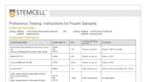
产品号#:
04100
18056
18056RF
产品名:
MethoCult™ H4100
Maes C et al. (MAY 2006)
The Journal of clinical investigation 116 5 1230--42
Placental growth factor mediates mesenchymal cell development, cartilage turnover, and bone remodeling during fracture repair.
Current therapies for delayed- or nonunion bone fractures are still largely ineffective. Previous studies indicated that the VEGF homolog placental growth factor (PlGF) has a more significant role in disease than in health. Therefore we investigated the role of PlGF in a model of semi-stabilized bone fracture healing. Fracture repair in mice lacking PlGF was impaired and characterized by a massive accumulation of cartilage in the callus,reminiscent of delayed- or nonunion fractures. PlGF was required for the early recruitment of inflammatory cells and the vascularization of the fracture wound. Interestingly,however,PlGF also played a role in the subsequent stages of the repair process. Indeed in vivo and in vitro findings indicated that PlGF induced the proliferation and osteogenic differentiation of mesenchymal progenitors and stimulated cartilage turnover by particular MMPs. Later in the process,PlGF was required for the remodeling of the newly formed bone by stimulating osteoclast differentiation. As PlGF expression was increased throughout the process of bone repair and all the important cell types involved expressed its receptor VEGFR-1,the present data suggest that PlGF is required for mediating and coordinating the key aspects of fracture repair. Therefore PlGF may potentially offer therapeutic advantages for fracture repair.
View Publication
Pirson L et al. (JUL 2006)
Stem cells (Dayton,Ohio) 24 7 1814--21
Despite inhibition of hematopoietic progenitor cell growth in vitro, the tyrosine kinase inhibitor imatinib does not impair engraftment of human CD133+ cells into NOD/SCIDbeta2mNull mice.
There is potential interest for combining allogeneic hematopoietic cell transplantation (HCT),and particularly allogeneic HCT with a nonmyeloablative regimen,to the tyrosine kinase inhibitor imatinib (Glivec; Novartis,Basel,Switzerland,http://www.novartis.com) in order to maximize anti-leukemic activity against Philadelphia chromosome-positive leukemias. However,because imatinib inhibits c-kit,the stem cell factor receptor,it could interfere with bone marrow engraftment. In this study,we examined the impact of imatinib on normal progenitor cell function. Imatinib decreased the colony-forming capacity of mobilized peripheral blood human CD133(+) cells but not that of long-term culture-initiating cells. Imatinib also decreased the proliferation of cytokine-stimulated CD133(+) cells but did not induce apoptosis of these cells. Expression of very late antigen (VLA)-4,VLA-5,and CXCR4 of CD133(+) cells was not modified by imatinib,but imatinib decreased the ability of CD133(+) cells to migrate. Finally,imatinib did not decrease engraftment of CD133(+) cells into irradiated nonobese diabetic/severe combined immunodeficient/beta2m(null) mice conditioned with 3 or 1 Gy total body irradiation. In summary,our results suggest that,despite inhibition of hematopoietic progenitor cell growth in vitro,imatinib does not interfere with hematopoietic stem cell engraftment.
View Publication
Miyake N et al. (MAR 2006)
Stem cells (Dayton,Ohio) 24 3 653--61
HOXB4-induced self-renewal of hematopoietic stem cells is significantly enhanced by p21 deficiency.
Enforced expression of the HOXB4 transcription factor and downregulation of p21(Cip1/Waf) (p21) can each independently increase proliferation of murine hematopoietic stem cells (HSCs). We asked whether the increase in HSC self-renewal generated by overexpression of HOXB4 is enhanced in p21-deficient HSCs. HOXB4 was overexpressed in hematopoietic cells from wild-type (wt) and p21-/- mice. Bone marrow (BM) cells were transduced with a retroviral vector expressing HOXB4 together with GFP (MIGB4),or a control vector containing GFP alone (MIG) and maintained in liquid culture for up to 11 days. At day 11 of the expansion culture,the number of primary CFU-GM (colony-forming unit granulocyte-macrophage) colonies and the repopulating ability were significantly increased in MIGB4 p21-/- BM (p21B4) cells compared with MIGB4-transduced wt BM (wtB4) cells. To test proliferation of HSCs in vivo,we performed competitive repopulation experiments and obtained significantly higher long-term engraftment of expanded p21B4 cells compared with wtB4 cells. The 5-day expansion of p21B4 HSCs generated 100-fold higher numbers of competitive repopulating units compared with wtMIG and threefold higher numbers compared with wtB4. The findings demonstrate that increased expression of HOXB4,in combination with suppression of p21 expression,could be a useful strategy for effective and robust expansion of HSCs.
View Publication

 EasySep™小鼠TIL(CD45)正选试剂盒


 沪公网安备31010102008431号