Grajales L et al. (APR 2010)
Journal of molecular and cellular cardiology 48 4 735--45
Delayed enrichment of mesenchymal cells promotes cardiac lineage and calcium transient development.
Bone marrow-derived mesenchymal stem cells (BM-MSCs) can be induced to differentiate into myogenic cells. Despite their potential,previous studies have not been successful in producing a high percentage of cardiac-like cells with a muscle phenotype. We hypothesized that cardiac lineage development in BM-MSC is related to cell passage,culture milieu,and enrichment for specific cell subtypes before and during differentiation. Our study demonstrated that Lin(-) BM-MSC at an intermediate passage (IP; P8-P12) expressed cardiac troponin T (cTnT) after 21 days in culture. Cardiac TnT expression was similar whether IP cells were differentiated in media containing 5-azacytidine+2% FBS (AZA; 14%) or 2% FBS alone (LS; 12%) and both were significantly higher than AZA+5% FBS. This expression was potentiated by first enriching for CD117/Sca-1 cells followed by differentiation (AZA,39% and LS,28%). A second sequential enrichment for the dihydropyridine receptor subunit alpha2delta1 (DHPR-alpha2) resulted in cardiac TnT expressed in 54% of cultured cells compared to 28% of cells after CD117/Sca-1(+) enrichment. Cells enriched for CD117/Sca-1 and subjected to differentiation displayed spontaneous intracellular Ca(2+) transients with an increase in transient frequency and a 60% decrease in the transient duration amplitude between days 14 and 29. In conclusion,IP CD117/Sca-1(+) murine BM-MSCs display robust cardiac muscle lineage development that can be induced independent of AZA but is diminished under higher serum concentrations. Furthermore,temporal changes in calcium kinetics commensurate with increased cTnT expression suggest progressive maturation of a cardiac muscle lineage. Enrichment with CD117/Sca-1 to establish lineage commitment followed by DHPR-alpha2 in lineage developing cells may enhance the therapeutic potential of these cells for transplantation.
View Publication
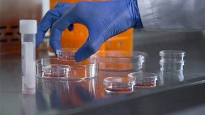
产品号#:
19771
产品名:
EasySep™ 小鼠间充质干/祖细胞富集试剂盒
Dí et al. (DEC 2010)
Cardiovascular research 88 3 502--11
Endothelial progenitor cells undergo an endothelial-to-mesenchymal transition-like process mediated by TGFbetaRI.
AIMS: Endothelial progenitor cells (EPC) have been shown to repair pulmonary endothelium,although they can also migrate into the arterial intima and differentiate into smooth muscle-like (mesenchymal) cells contributing to intimal hyperplasia. The molecular mechanisms by which this process proceeds have not been fully elucidated. Here,we study whether genes involved in the endothelial-to-mesenchymal transition (EnMT) may contribute to the mesenchymal phenotype acquisition of EPC and we evaluate whether transforming growth factor β1 (TGFβ1) is involved in this process. METHODS AND RESULTS: Our results show that co-culture of EPC with smooth muscle cells (SMC) increases the expression of the mesenchymal cell markers α-smooth muscle actin,sm22-α,and myocardin,and decreases the expression of the endothelial cell marker CD31. In the same conditions,we also observed a concomitant increase in the gene expression of the EnMT-related transcription factors: slug,snail,zeb1,and endothelin-1. This indicates that mesenchymal phenotype acquisition occurred through an EnMT-like process. Inhibition of TGFβ receptor I (TGFβRI) downregulated snail gene expression,blocked the EnMT,and facilitated the differentiation of EPC to the endothelial cell lineage. Furthermore,TGFβRI inhibition decreased migration of EPC stimulated by SMC without affecting their functionality and adhesion capacity. CONCLUSION: These results indicate that EPC may differentiate into SMC-like cells through an EnMT-like process and that TGFβI plays an important role in the fate of EPC.
View Publication
Lee J-H et al. (JUL 2005)
Experimental cell research 307 1 174--82
Contribution of human bone marrow stem cells to individual skeletal myotubes followed by myogenic gene activation.
Much attention is focused on characterizing the contribution of bone marrow (BM)-derived cells to regenerating skeletal muscle,fuelled by hopes for stem cell-mediated therapy of muscle degenerative diseases. Though physical integration of BM stem cells has been well documented,little evidence of functional commitment to myotube phenotype has been reported. This is due to the innate difficulty in distinguishing gene products derived from donor versus host nuclei. Here,we demonstrate that BM-derived stem cells contribute via gene expression following incorporation to skeletal myotubes. By co-culturing human BM-derived mesenchymal stem cells (MSC) with mouse skeletal myoblasts,physical incorporation was observed by genetic lineage tracing and species-specific immunofluorescence. We used a human-specific antibody against the intermediate filament protein nestin,a marker of regenerating skeletal muscle,to identify functional contribution of MSC to myotube formation. Although nestin expression was never detected in MSC,human-specific expression was detected in myotubes that also contained MSC-derived nuclei. This induction of gene expression following myotube integration suggests that bone marrow-derived stem cells can reprogram and functionally contribute to the muscle cell phenotype. We propose that this model of myogenic commitment may provide the means to further characterize functional reprogramming of MSC to skeletal muscle.
View Publication
产品号#:
05401
15128
15168
产品名:
MesenCult™ MSC 基础培养基(人)
RosetteSep™人间充质干细胞富集抗体混合物
RosetteSep™人间充质干细胞富集抗体混合物
Maes C et al. (MAY 2006)
The Journal of clinical investigation 116 5 1230--42
Placental growth factor mediates mesenchymal cell development, cartilage turnover, and bone remodeling during fracture repair.
Current therapies for delayed- or nonunion bone fractures are still largely ineffective. Previous studies indicated that the VEGF homolog placental growth factor (PlGF) has a more significant role in disease than in health. Therefore we investigated the role of PlGF in a model of semi-stabilized bone fracture healing. Fracture repair in mice lacking PlGF was impaired and characterized by a massive accumulation of cartilage in the callus,reminiscent of delayed- or nonunion fractures. PlGF was required for the early recruitment of inflammatory cells and the vascularization of the fracture wound. Interestingly,however,PlGF also played a role in the subsequent stages of the repair process. Indeed in vivo and in vitro findings indicated that PlGF induced the proliferation and osteogenic differentiation of mesenchymal progenitors and stimulated cartilage turnover by particular MMPs. Later in the process,PlGF was required for the remodeling of the newly formed bone by stimulating osteoclast differentiation. As PlGF expression was increased throughout the process of bone repair and all the important cell types involved expressed its receptor VEGFR-1,the present data suggest that PlGF is required for mediating and coordinating the key aspects of fracture repair. Therefore PlGF may potentially offer therapeutic advantages for fracture repair.
View Publication
产品号#:
03534
03334
03434
03444
18753
18753RF
产品名:
MethoCult™ GF M3534
MethoCult™ M3334
MethoCult™ GF M3434
MethoCult™ GF M3434
Muguruma Y et al. (MAR 2006)
Blood 107 5 1878--87
Reconstitution of the functional human hematopoietic microenvironment derived from human mesenchymal stem cells in the murine bone marrow compartment.
Hematopoiesis is maintained by specific interactions between both hematopoietic and nonhematopoietic cells. Whereas hematopoietic stem cells (HSCs) have been extensively studied both in vitro and in vivo,little is known about the in vivo characteristics of stem cells of the nonhematopoietic component,known as mesenchymal stem cells (MSCs). Here we have visualized and characterized human MSCs in vivo following intramedullary transplantation of enhanced green fluorescent protein-marked human MSCs (eGFP-MSCs) into the bone marrow (BM) of nonobese diabetic/severe combined immunodeficiency (NOD/SCID) mice. Between 4 to 10 weeks after transplantation,eGFP-MSCs that engrafted in murine BM integrated into the hematopoietic microenvironment (HME) of the host mouse. They differentiated into pericytes,myofibroblasts,BM stromal cells,osteocytes in bone,bone-lining osteoblasts,and endothelial cells,which constituted the functional components of the BM HME. The presence of human MSCs in murine BM resulted in an increase in functionally and phenotypically primitive human hematopoietic cells. Human MSC-derived cells that reconstituted the HME appeared to contribute to the maintenance of human hematopoiesis by actively interacting with primitive human hematopoietic cells.
View Publication
产品号#:
04034
04044
产品名:
MethoCult™ H4034 Optimum
MethoCult™ H4034 Optimum
Zhang H et al. (NOV 2005)
American journal of physiology. Heart and circulatory physiology 289 5 H2089--96
Increasing donor age adversely impacts beneficial effects of bone marrow but not smooth muscle myocardial cell therapy.
We evaluated the impact of donor age on the efficacy of myocardial cellular therapy for ischemic cardiomyopathy. Characteristics of smooth muscle cells (SMC),bone marrow stromal cells (MSCs),and skeletal muscle cells (SKMCs) from young,adult,and old rats were compared in vitro. Three weeks after coronary ligation,3.5 million SMCs (n = 11) or MSCs (n = 9) from old syngenic rats or culture medium (n = 6) were injected into the ischemic region. Five weeks after implantation,cardiac function was assessed by echocardiography and the Langendorff apparatus. In the in vitro study,the numbers and proliferation of MSCs from fresh bone marrow and SKMCs from fresh tissue but not SMCs were markedly diminished in old animals (P textless 0.05 both groups). SKMCs from old animals did not reach confluence. After treatment with 5-azacytidine (azacitidine),the myogenic potential of old MSCs was decreased compared with young MSCs. In the in vivo study,both SMC and MSC transplantation induced significant angiogenesis compared with media injections (P textless 0.05 both groups). Transplantation of SMCs but not MSCs prevented scar thinning (P = 0.03) and improved ejection fraction and fractional shortening (P textless 0.05). Load-independent indices of cardiac function in a Langendorff preparation confirmed improved function in the aged SMC group (P = 0.01) but not in the MSC group compared with the control group. In conclusion,donor age adversely impacts the efficacy of cellular therapy for myocardial regeneration and is cell-type dependent. SMCs from old donors retain their ability to improve cardiac function after implantation into ischemic myocardium.
View Publication
产品号#:
05501
05502
产品名:
Mendelson A et al. (OCT 2011)
FASEB journal : official publication of the Federation of American Societies for Experimental Biology 25 10 3496--504
Chondrogenesis by chemotactic homing of synovium, bone marrow, and adipose stem cells in vitro.
Cell transplantation has been well explored for cartilage regeneration. We recently showed that the entire articular surface of a synovial joint can regenerate by endogenous cell homing and without cell transplantation. However,the sources of endogenous cells that regenerate articular cartilage remain elusive. Here,we studied whether cytokines not only chemotactically recruit adipose stem cells (ASCs),mesenchymal stem cells (MSCs),and synovium stem cells (SSCs) but also induce chondrogenesis of the recruited cells. Recombinant human transforming growth factor-β3 (TGF-β3; 100 ng) and/or recombinant human stromal derived factor-1β (SDF-1β; 100 ng) was control released into an acellular collagen sponge cube with underlying ASCs,MSCs,or SSCs in monolayer culture. Although all cell types randomly migrated into the acellular collagen sponge cube,TGF-β3 and/or SDF-1β recruited significantly more cells than the cytokine-free control group. In 6 wk,TGF-β3 alone recruited substantial numbers of ASCs (558±65) and MSCs (302±52),whereas codelivery of TGF-β3 and SDF-1β was particularly chemotactic to SSCs (400±120). Proliferation of the recruited cells accounted for some,but far from all,of the observed cellularity. TGF-β3 and SDF-1β codelivery induced significantly higher aggrecan gene expression than the cytokine-free group for ASCs,MSCs,and SSCs. Type II collagen gene expression was also significantly higher for ASCs and SSCs by SDF-1 and TGF-β3 codelivery. Remarkably,the expression of aggrecan and type II collagen was detected among all cell types. Thus,homing of multiple stem/progenitor cell populations may potentially serve as an alternative or adjunctive approach to cell transplantation for cartilage regeneration.
View Publication
产品号#:
05401
05402
05411
产品名:
MesenCult™ MSC 基础培养基(人)
MesenCult™ MSC 刺激补充剂(人)
MesenCult™ 增殖试剂盒(人)
Tauchmanovà et al. (MAY 2003)
Cancer 97 10 2453--61
Avascular necrosis in long-term survivors after allogeneic or autologous stem cell transplantation: a single center experience and a review.
BACKGROUND: The most debilitating skeletal complication of stem cell transplantation (SCT) is avascular necrosis (AVN). METHODS: Two hundred seven consecutive patients were evaluated prospectively for AVN. They survived disease free for more than 180 days after autologous or allogeneic SCT for hematologic malignancies. The diagnosis of AVN in suspicious cases was confirmed by magnetic resonance imaging. Possible correlations with treatments,bone mineral density (BMD),graft versus host disease (GVHD),and in vitro growth of fibroblast progenitors were investigated. Bone mineral density was evaluated by dual-energy X-ray absorptiometry in 100 transplanted patients,and the in vitro growth of fibroblast progenitors was monitored by a fibroblast colony-forming unit (CFU-F) assay in 30 patients after allogeneic SCT. RESULTS: Twelve patients developed AVN 3-114 months (median,26 months) following SCT: 10 (10%) after allogeneic SCT and 2 (1.9%) after autologous SCT (P = 0.04). Twenty-five joints were affected by AVN. All patients had femoral head involvement,which was managed with hip replacement in six of them. All but one patient who developed AVN after allogeneic SCT suffered from chronic GVHD (cGVHD). Avascular necrosis occurred 1-4 months after exacerbation or progression of cGVHD. Cumulative dose of steroids was similar in both SCT groups (including steroids given pretransplant for the basic disease),whereas treatment duration was significantly longer in the allogeneic SCT group. Avascular necrosis was related to the decreased number of bone marrow CFU-F colonies in vitro,but not to BMD values. CONCLUSIONS: Avascular necrosis is a skeletal complication that occurs more often after allogeneic than after autologous SCT. Occurrence of AVN symptoms after clinical follow-up of cGVHD suggests that cGVHD requiring long-term steroid therapy is one of the main risk factors for AVN. Avascular necrosis may be facilitated by a severe deficit in the repopulating capacity of bone marrow stromal stem cells after SCT.
View Publication
产品号#:
05401
05402
05411
产品名:
MesenCult™ MSC 基础培养基(人)
MesenCult™ MSC 刺激补充剂(人)
MesenCult™ 增殖试剂盒(人)
Lapter S et al. (MAR 2007)
Stem cells (Dayton,Ohio) 25 3 761--70
Structure and implied functions of truncated B-cell receptor mRNAs in early embryo and adult mesenchymal stem cells: Cdelta replaces Cmu in mu heavy chain-deficient mice.
Stem cells exhibit a promiscuous gene expression pattern. We show herein that the early embryo and adult MSCs express B-cell receptor component mRNAs. To examine possible bearings of these genes on the expressing cells,we studied immunoglobulin mu chain-deficient mice. Pregnant mu chain-deficient females were found to produce a higher percentage of defective morulae compared with control females. Structure analysis indicated that the mu mRNA species found in embryos and in mesenchyme consist of the constant region of the mu heavy chain that encodes a recombinant 50-kDa protein. In situ hybridization localized the constant mu gene expression to loose mesenchymal tissues within the day-12.5 embryo proper and the yolk sac. In early embryo and in adult mesenchyme from mu-deficient mice,delta replaced mu chain,implying a possible requirement of these alternative molecules for embryo development and mesenchymal functions. Indeed,overexpression of the mesenchymal-truncated mu heavy chain in 293T cells resulted in specific subcellular localization and in G(1) growth arrest. The lack of such occurrence following overexpression of a complete,rearranged form of mu chain suggests that the mesenchymal version of this mRNA may possess unique functions.
View Publication

 EasySep™小鼠TIL(CD45)正选试剂盒


 沪公网安备31010102008431号