Schmuck EG et al. (MAR 2014)
Cardiovascular engineering and technology 5 1 119--131
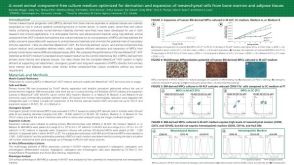
Cardiac fibroblast-derived 3D extracellular matrix seeded with mesenchymal stem cells as a novel device to transfer cells to the ischemic myocardium.
PURPOSE Demonstrate a novel manufacturing method to generate extracellular matrix scaffolds from cardiac fibroblasts (CF-ECM) as a therapeutic mesenchymal stem cell-transfer device. MATERIALS AND METHODS Rat CF were cultured at high-density (˜1.6×10(5)/cm(2)) for 10-14 days. Cell sheets were removed from the culture dish by incubation with EDTA and decellularized with water and peracetic acid. CF-ECM was characterized by mass spectrometry,immunofluorescence and scanning electron microscopy. CF-ECM seeded with human embryonic stem cell derived mesenchymal stromal cells (hEMSCs) were transferred into a mouse myocardial infarction model. 48 hours later,mouse hearts were excised and examined for CF-ECM scaffold retention and cell transfer. RESULTS CF-ECM scaffolds are composed of fibronectin (82%),collagens type I (13%),type III (3.4%),type V (0.2%),type II (0.1%) elastin (1.3%) and 18 non-structural bioactive molecules. Scaffolds remained intact on the mouse heart for 48 hours without the use of sutures or glue. Identified hEMSCs were distributed from the epicardium to the endocardium. CONCLUSIONS High density cardiac fibroblast culture can be used to generate CF-ECM scaffolds. CF-ECM scaffolds seeded with hEMSCs can be maintained on the heart without suture or glue. hEMSC are successfully delivered throughout the myocardium.
View Publication
Perez JE et al. (FEB 2017)
Nanotechnology 28 5 55703
Mesenchymal stem cells cultured on magnetic nanowire substrates.
Stem cells have been shown to respond to extracellular mechanical stimuli by regulating their fate through the activation of specific signaling pathways. In this work,an array of iron nanowires (NWs) aligned perpendicularly to the surface was fabricated by pulsed electrodepositon in porous alumina templates followed by a partial removal of the alumina to reveal 2-3 μm of the NWs. This resulted in alumina substrates with densely arranged NWs of 33 nm in diameter separated by 100 nm. The substrates were characterized by scanning electron microscopy (SEM) energy dispersive x-ray analysis and vibrating sample magnetometer. The NW array was then used as a platform for the culture of human mesenchymal stem cells (hMSCs). The cells were stained for the cell nucleus and actin filaments,as well as immuno-stained for the focal adhesion protein vinculin,and then observed by fluorescence microscopy in order to characterize their spreading behavior. Calcein AM/ethidium homodimer-1 staining allowed the determination of cell viability. The interface between the cells and the NWs was studied using SEM. Results showed that hMSCs underwent a re-organization of actin filaments that translated into a change from an elongated to a spherical cell shape. Actin filaments and vinculin accumulated in bundles,suggesting the attachment and formation of focal adhesion points of the cells on the NWs. Though the overall number of cells attached on the NWs was lower compared to the control,the attached cells maintained a high viability (>90%) for up to 6 d. Analysis of the interface between the NWs and the cells confirmed the re-organization of F-actin and revealed the adhesion points of the cells on the NWs. Additionally,a net of filopodia surrounded each cell,suggesting the probing of the array to find additional adhesion points. The cells maintained their round shape for up to 6 d of culture. Overall,the NW array is a promising nanostructured platform for studying and influencing hMSCs differentiation.
View Publication
Sessarego N et al. (MAR 2008)
Haematologica 93 3 339--46
Multipotent mesenchymal stromal cells from amniotic fluid: solid perspectives for clinical application.
BACKGROUND: Mesenchymal stromal cells are multipotent cells considered to be of great promise for use in regenerative medicine. However,the cell dose may be a critical factor in many clinical conditions and the yield resulting from the ex vivo expansion of mesenchymal stromal cells derived from bone marrow may be insufficient. Thus,alternative sources of mesenchymal stromal cells need to be explored. In this study,mesenchymal stromal cells were successfully isolated from second trimester amniotic fluid and analyzed for chromosomal stability to validate their safety for potential utilization as a cell therapy product. DESIGN AND METHODS: Mesenchymal stromal cells were expanded up to the sixth passage starting from amniotic fluid using different culture conditions to optimize large-scale production. RESULTS: The highest number of mesenchymal stromal cells derived from amniotic fluid was reached at a low plating density; in these conditions the expansion of mesenchymal stromal cells from amniotic fluid was significantly greater than that of adult bone marrow-derived mesenchymal stromal cells. Mesenchymal stromal cells from amniotic fluid represent a relatively homogeneous population of immature cells with immunosuppressive properties and extensive proliferative potential. Despite their high proliferative capacity in culture,we did not observe any karyotypic abnormalities or transformation potential in vitro nor any tumorigenic effect in vivo. CONCLUSIONS: Fetal mesenchymal stromal cells can be extensively expanded from amniotic fluid,showing no karyotypic abnormalities or transformation potential in vitro and no tumorigenic effect in vivo. They represent a relatively homogeneous population of immature mesenchymal stromal cells with long telomeres,immunosuppressive properties and extensive proliferative potential. Our results indicate that amniotic fluid represents a rich source of mesenchymal stromal cells suitable for banking to be used when large amounts of cells are required.
View Publication
产品号#:
05401
05402
05411
产品名:
MesenCult™ MSC 基础培养基(人)
MesenCult™ MSC刺激添加物(人)
MesenCult™ 增殖试剂盒(人)
Schumann P et al. (SEP 2009)
Microvascular research 78 2 180--90
Consequences of seeded cell type on vascularization of tissue engineering constructs in vivo.
Implantation of tissue engineering constructs is a promising technique to reconstruct injured tissue. However,after implantation the nutrition of the constructs is predominantly restricted to vascularization. Since cells possess distinct angiogenic potency,we herein assessed whether scaffold vitalization with different cell types improves scaffold vascularization. 32 male balb/c mice received a dorsal skinfold chamber. Angiogenesis,microhemodynamics,leukocyte-endothelial cell interaction and microvascular permeability induced in the host tissue after implantation of either collagen coated poly (L-lactide-co-glycolide) (PLGA) scaffolds (group 4),additionally seeded with osteoblast-like cells (OLCs,group 1),bone marrow mesenchymal stem cells (bmMSCs,group 2) or a combination of OLCs and bmMSCs (group 3) were analyzed repetitively over 14 days using intravital fluorescence microscopy. Apart from a weak inflammatory response in all groups,vascularization was found distinctly accelerated in vitalized scaffolds,indicated by a significantly increased microvascular density (day 6,group 1: 202+/-15 cm/cm(2),group 2: 202+/-12 cm/cm(2),group 3: 194+/-8 cm/cm(2)),when compared with controls (group 4: 72+/-5 cm/cm(2)). This acceleration was independent from the seeded cell type. Immunohistochemistry revealed in vivo VEGF expression in close vicinity to the seeded OLCs and bmMSCs. Therefore,the observed lack of cell type confined differences in the vascularization process suggests that the accelerated vascularization of vitalized scaffolds is VEGF-related rather than dependent on the potential of bmMSCs to differentiate into specific vascular cells.
View Publication
产品号#:
05501
05502
产品名:
Feng Y et al. (SEP 2010)
Progress in biophysics and molecular biology 103 1 148--56
Unique biomechanical interactions between myeloma cells and bone marrow stroma cells.
We observed that BMSCs (bone marrow stromal cells) from myeloma patients (myeloma BMSCs) were significantly stiffer than control BMSCs using a cytocompression device. The stiffness of myeloma BMSCs and control BMSCs was further increased upon priming by myeloma cells. Additionally,myeloma cells became stiffer when primed by myeloma BMSCs. The focal adhesion kinase activity of myeloma cells was increased when cells were on stiffer collagen gels and on myeloma BMSCs. This change in myeloma stiffness is associated with increased colony formation of myeloma cells and FAK activation when co-cultured with stiffer myeloma BMSCs or stiffer collagen. Additionally,stem cells of RPMI8226 cells became stiffer after priming by myeloma BMSCs,with concomitant increases of stem cell colony formation. These results suggest the presence of a mechanotransduction loop between myeloma cells and myeloma BMSCs to increase the stiffness of both types of cells via FAK activation. The increase of stiffness may in turn support the growth of myeloma cells and myeloma stem cells.
View Publication
产品号#:
05401
05402
05411
产品名:
MesenCult™ MSC 基础培养基(人)
MesenCult™ MSC刺激添加物(人)
MesenCult™ 增殖试剂盒(人)
Thirumala S et al. (JUL 2009)
Organogenesis 5 3 143--54
Clinical grade adult stem cell banking.
There has been a great deal of scientific interest recently generated by the potential therapeutic applications of adult stem cells in human care but there are several challenges regarding quality and safety in clinical applications and a number of these challenges relate to the processing and banking of these cells ex-vivo. As the number of clinical trials and the variety of adult cells used in regenerative therapy increases,safety remains a primary concern. This has inspired many nations to formulate guidelines and standards for the quality of stem cell collection,processing,testing,banking,packaging and distribution. Clinically applicable cryopreservation and banking of adult stem cells offers unique opportunities to advance the potential uses and widespread implementation of these cells in clinical applications. Most current cryopreservation protocols include animal serum proteins and potentially toxic cryoprotectant additives (CPAs) that prevent direct use of these cells in human therapeutic applications. Long term cryopreservation of adult stem cells under good manufacturing conditions using animal product free solutions is critical to the widespread clinical implementation of ex-vivo adult stem cell therapies. Furthermore,to avoid any potential cryoprotectant related complications,reduced CPA concentrations and efficient post-thaw washing to remove CPA are also desirable. The present review focuses on the current strategies and important aspects of adult stem cell banking for clinical applications. These include current good manufacturing practices (cGMPs),animal protein free freezing solutions,cryoprotectants,freezing & thawing protocols,viability assays,packaging and distribution. The importance and benefits of banking clinical grade adult stem cells are also discussed.
View Publication
产品号#:
07930
07931
07940
07955
07956
07959
07954
100-1061
07952
产品名:
CryoStor® CS10
CryoStor® CS10
CryoStor® CS10
CryoStor® CS10
CryoStor® CS10
CryoStor® CS10
CryoStor® CS10
Rasheed ZA et al. (MAR 2010)
Journal of the National Cancer Institute 102 5 340--51
Prognostic significance of tumorigenic cells with mesenchymal features in pancreatic adenocarcinoma.
BACKGROUND: Specific populations of highly tumorigenic cells are thought to exist in many human tumors,including pancreatic adenocarcinoma. However,the clinical significance of these tumor-initiating (ie,cancer stem) cells remains unclear. Aldehyde dehydrogenase (ALDH) activity can identify tumor-initiating cells and normal stem cells from several human tissues. We examined the prognostic significance and functional features of ALDH expression in pancreatic adenocarcinoma. METHODS: ALDH expression was analyzed by immunohistochemistry in 269 primary surgical specimens of pancreatic adenocarcinoma and examined for association with clinical outcomes and in paired primary tumors and metastatic lesions from eight pancreatic cancer patients who had participated in a rapid autopsy program. The clonogenic growth potential of ALDH-positive pancreatic adenocarcinoma cells was assessed in vitro by a colony formation assay and by tumor growth in immunodeficient mice (10-14 mice per group). Mesenchymal features of ALDH-positive pancreatic tumor cells were examined by using quantitative reverse transcription-polymerase chain reaction and an in vitro cell invasion assay. Gene expression levels and the invasive potential of ADLH-positive pancreatic cancer cells relative to the bulk cell population were examined by reverse transcription-polymerase chain reaction and an in vitro invasion assays,respectively. All statistical tests were two-sided. RESULTS: ALDH-positive tumor cells were detected in 90 of the 269 primary surgical specimens,and their presence was associated with worse survival (median survival for patients with ALDH-positive vs ALDH-negative tumors: 14 vs 18 months,hazard ratio of death = 1.28,95% confidence interval = 1.02 to 1.68,P = .05). Six (75%) of the eight patients with matched primary and metastatic tumor samples had ALDH-negative primary tumors,and in four (67%) of these six patients,the matched metastatic lesions (located in liver and lung) contained ALDH-positive cells. ALDH-positive cells were approximately five- to 11-fold more clonogenic in vitro and in vivo compared with unsorted or ALHD-negative cells,expressed genes consistent with a mesenchymal state,and had in vitro migratory and invasive potentials that were threefold greater than those of unsorted cells. CONCLUSIONS: ALDH expression marks pancreatic cancer cells that have stem cell and mesenchymal features. The enhanced clonogenic growth and migratory properties of ALDH-positive pancreatic cancer cells suggest that they play a key role in the development of metastatic disease that negatively affects the overall survival of patients with pancreatic adenocarcinoma.
View Publication

 EasySep™小鼠TIL(CD45)正选试剂盒


 沪公网安备31010102008431号