Valencic E et al. (APR 2010)
Cytotherapy 12 2 154--60
The immunosuppressive effect of Wharton's jelly stromal cells depends on the timing of their licensing and on lymphocyte activation.
BACKGROUND: Mesenchymal stromal cells (MSC) have been proven to have potent immunosuppressive action and hence have been proposed for the treatment of severe Graft Versus Host Disease. However,in most models,MSC were added at the same time of lymphocyte stimulation,which is quite different from what occurs in vivo. AIMS: To investigate how the timing of lymphocyte activation and the exposure to activation-related cytokines (licensing) can influence the immunosuppressive action of Wharton's jelly stromal cells (WJSC). METHODS: WJSC,licensed or not with activation-related cytokines,were added lymphocytes the same time or 24 hours after their stimulation with phytohaemoagglutinin. Proliferation of lymphocytes and cytokines production was measured after three days co-culture. RESULTS: Lymphocytes stimulated in the presence of WJSC displayed a dramatic decrease in proliferation and production of cytokines,in spite of normal expression of activation markers. The suppression was weakened when targeted lymphocytes were seperated by a membrane and partially rescued by the addition of exogenous l-tryptophan,suggesting a major role for indoleamine 2,3-dioxigenase with a probable paracrine effect. Licensing of WJSC increased the immunosuppressive effect,in both contact and non-contact settings. The timing of WJSC licensing was crucial for the immunosuppressive action. Lymphocytes pre-stimulated alone for 24 h,and added afterwards to non-licensed WJSC,showed normal or even increased proliferation. On the other hand,their proliferation was strongly inhibited by licensed WJSC. CONCLUSIONS: WJSC have a potent immunosuppressive function best realized with direct contact,and increased by licensing signals before and during lymphocyte stimulation. Our results could contribute to the set up of new WJSC-based therapies for severe autoimmuno disorders.
View Publication
产品号#:
产品名:
Nakamura Y et al. (SEP 2010)
Blood 116 9 1422--32
Isolation and characterization of endosteal niche cell populations that regulate hematopoietic stem cells.
The endosteal niche is critical for the maintenance of hematopoietic stem cells (HSCs). However,it consists of a heterogeneous population in terms of differentiation stage and function. In this study,we characterized endosteal cell populations and examined their ability to maintain HSCs. Bone marrow endosteal cells were subdivided into immature mesenchymal cell-enriched ALCAM(-)Sca-1(+) cells,osteoblast-enriched ALCAM(+)Sca-1(-),and ALCAM(-)Sca-1(-) cells. We found that all 3 fractions maintained long-term reconstitution (LTR) activity of HSCs in an in vitro culture. In particular,ALCAM(+)Sca-1(-) cells significantly enhanced the LTR activity of HSCs by the up-regulation of homing- and cell adhesion-related genes in HSCs. Microarray analysis showed that ALCAM(-)Sca-1(+) fraction highly expressed cytokine-related genes,whereas the ALCAM(+)Sca-1(-) fraction expressed multiple cell adhesion molecules,such as cadherins,at a greater level than the other fractions,indicating that the interaction between HSCs and osteoblasts via cell adhesion molecules enhanced the LTR activity of HSCs. Furthermore,we found an osteoblastic marker(low/-) subpopulation in ALCAM(+)Sca-1(-) fraction that expressed cytokines,such as Angpt1 and Thpo,and stem cell marker genes. Altogether,these data suggest that multiple subsets of osteoblasts and mesenchymal progenitor cells constitute the endosteal niche and regulate HSCs in adult bone marrow.
View Publication
产品号#:
03434
03444
产品名:
MethoCult™ GF M3434
MethoCult™ GF M3434
Yañ et al. (NOV 2010)
Experimental cell research 316 19 3109--23
Prostaglandin E2 plays a key role in the immunosuppressive properties of adipose and bone marrow tissue-derived mesenchymal stromal cells.
Mesenchymal stromal cells (MSCs) have important immunosuppressive properties,but the mechanisms and soluble factors involved in these effects remain unclear. We have studied prostaglandin-E2 (PGE2) as a possible candidate implied in adipose tissue-derived MSCs (Ad-MSCs) immunosuppressive properties over dendritic cells and T lymphocytes,compared to bone marrow derived MSCs (BM-MSCs). We found that both MSCs inhibited the maturation of myeloid-DCs and plasmocytoid-DCs. High levels of PGE2 were detected in DCs/MSCs co-cultures. Its blockade with indomethacin (IDM) allowed plasmocytoid-DCs but not myeloid-DCs maturation. Additionally,high levels of PGE2 were found in co-cultures in which Ad-MSCs or BM-MSCs inhibited activated T cells proliferation and pro-inflammatory cytokines production. PGE2 blockade by IDM preserved T lymphocytes proliferation but did not restore the pro-inflammatory cytokines secretion. However,an increased expression of transcription factors and cytokines genes involved in the Th1/Th2 differentiation pathway was detected in the T cells co-cultured with Ad-MSCs,but not with BM-MSCs. In conclusion,we propose that PGE2 is a soluble factor mediating most of the immunosuppressive effects of Ad-MSCs and BM-MSCs over p-DCs maturation and activated T lymphocytes proliferation and cytokine secretion.
View Publication
产品号#:
05401
05402
05411
产品名:
MesenCult™ MSC 基础培养基(人)
MesenCult™ MSC 刺激补充剂(人)
MesenCult™ 增殖试剂盒(人)
Rubin MR et al. (JAN 2011)
The Journal of clinical endocrinology and metabolism 96 1 176--86
Parathyroid hormone stimulates circulating osteogenic cells in hypoparathyroidism.
CONTEXT: The osteoanabolic properties of PTH may be due to increases in the number and maturity of circulating osteogenic cells. Hypoparathyroidism is a useful clinical model because this hypothesis can be tested by administering PTH. OBJECTIVE: The objective of the study was to characterize circulating osteogenic cells in hypoparathyroid subjects during 12 months of PTH (1-84) administration. DESIGN: Osteogenic cells were characterized using flow cytometry and antibodies against osteocalcin,an osteoblast-specific protein product,and stem cell markers CD34 and CD146. Changes in bone formation from biochemical markers and quadruple-labeled transiliac crest bone biopsies (0 and 3 month time points) were correlated with measurements of circulating osteogenic cells. SETTING: The study was conducted at a clinical research center. PATIENTS: Nineteen control and 19 hypoparathyroid patients were included in the study. INTERVENTION: Intervention included the administration of PTH (1-84). RESULTS: Osteocalcin-positive cells were lower in hypoparathyroid subjects than controls (0.7 ± 0.1 vs. 2.0 ± 0.1%; P textless 0.0001),with greater coexpression of the early cell markers CD34 and CD146 among the osteocalcin-positive cells in the hypoparathyroid subjects (11.0 ± 1.0 vs. 5.6 ± 0.7%; P textless 0.001). With PTH (1-84) administration,the number of osteogenic cells increased 3-fold (P textless 0.0001),whereas the coexpression of the early cell markers CD34 and CD146 decreased. Increases in osteogenic cells correlated with circulating and histomorphometric indices of osteoblast function: N-terminal propeptide of type I procollagen (R(2) = 0.4,P ≤ 0.001),bone-specific alkaline phosphatase (R(2) = 0.3,P textless 0.001),osteocalcin (R(2) = 0.4,P textless 0.001),mineralized perimeter (R(2) = 0.5,P textless 0.001),mineral apposition rate (R(2) = 0.4,P = 0.003),and bone formation rate (R(2) = 0.5,P textless 0.001). CONCLUSIONS: It is likely that PTH stimulates bone formation by stimulating osteoblast development and maturation. Correlations between circulating osteogenic cells and histomorphometric indices of bone formation establish that osteoblast activity is being identified by this methodology.
View Publication
Wu X et al. (APR 2011)
The Journal of biological chemistry 286 15 13512--21
p85alpha regulates osteoblast differentiation by cross-talking with the MAPK pathway.
Class IA phosphoinositide 3-kinase (PI3K) is involved in regulating many cellular functions including cell growth,proliferation,cell survival,and differentiation. The p85 regulatory subunit is a critical component of the PI3K signaling pathway. Mesenchymal stem cells (MSC) are multipotent cells that can be differentiated into osteoblasts (OBs),adipocytes,and chondrocytes under defined culture conditions. To determine whether p85α subunit of PI3K affects biological functions of MSCs,bone marrow-derived wild type (WT) and p85α-deficient (p85α(-/-)) cells were employed in this study. Increased cell growth,higher proliferation rate and reduced number of senescent cells were observed in MSCs lacking p85α compare with WT MSCs as evaluated by CFU-F assay,thymidine incorporation assay,and β-galactosidase staining,respectively. These functional changes are associated with the increased cell cycle,increased expression of cyclin D,cyclin E,and reduced expression of p16 and p19 in p85α(-/-) MSCs. In addition,a time-dependent reduction in alkaline phosphatase (ALP) activity and osteocalcin mRNA expression was observed in p85α(-/-) MSCs compared with WT MSCs,suggesting impaired osteoblast differentiation due to p85α deficiency in MSCs. The impaired p85α(-/-) osteoblast differentiation was associated with increased activation of Akt and MAPK. Importantly,bone morphogenic protein 2 (BMP2) was able to intensify the differentiation of osteoblasts derived from WT MSCs,whereas this process was significantly impaired as a result of p85α deficiency. Addition of LY294002,a PI3K inhibitor,did not alter the differentiation of osteoblasts in either genotype. However,application of PD98059,a Mek/MAPK inhibitor,significantly enhanced osteoblast differentiation in WT and p85α(-/-) MSCs. These results suggest that p85α plays an essential role in osteoblast differentiation from MSCs by repressing the activation of MAPK pathway.
View Publication
产品号#:
05501
05502
产品名:
Mendelson A et al. (OCT 2011)
FASEB journal : official publication of the Federation of American Societies for Experimental Biology 25 10 3496--504
Chondrogenesis by chemotactic homing of synovium, bone marrow, and adipose stem cells in vitro.
Cell transplantation has been well explored for cartilage regeneration. We recently showed that the entire articular surface of a synovial joint can regenerate by endogenous cell homing and without cell transplantation. However,the sources of endogenous cells that regenerate articular cartilage remain elusive. Here,we studied whether cytokines not only chemotactically recruit adipose stem cells (ASCs),mesenchymal stem cells (MSCs),and synovium stem cells (SSCs) but also induce chondrogenesis of the recruited cells. Recombinant human transforming growth factor-β3 (TGF-β3; 100 ng) and/or recombinant human stromal derived factor-1β (SDF-1β; 100 ng) was control released into an acellular collagen sponge cube with underlying ASCs,MSCs,or SSCs in monolayer culture. Although all cell types randomly migrated into the acellular collagen sponge cube,TGF-β3 and/or SDF-1β recruited significantly more cells than the cytokine-free control group. In 6 wk,TGF-β3 alone recruited substantial numbers of ASCs (558±65) and MSCs (302±52),whereas codelivery of TGF-β3 and SDF-1β was particularly chemotactic to SSCs (400±120). Proliferation of the recruited cells accounted for some,but far from all,of the observed cellularity. TGF-β3 and SDF-1β codelivery induced significantly higher aggrecan gene expression than the cytokine-free group for ASCs,MSCs,and SSCs. Type II collagen gene expression was also significantly higher for ASCs and SSCs by SDF-1 and TGF-β3 codelivery. Remarkably,the expression of aggrecan and type II collagen was detected among all cell types. Thus,homing of multiple stem/progenitor cell populations may potentially serve as an alternative or adjunctive approach to cell transplantation for cartilage regeneration.
View Publication
产品号#:
05401
05402
05411
产品名:
MesenCult™ MSC 基础培养基(人)
MesenCult™ MSC 刺激补充剂(人)
MesenCult™ 增殖试剂盒(人)
Keller GM (DEC 1995)
Current opinion in cell biology 7 6 862--9
In vitro differentiation of embryonic stem cells.
Under appropriate conditions in culture,embryonic stem cells will differentiate and form embryoid bodies that have been shown to contain cells of the hematopoietic,endothelial,muscle and neuronal lineages. Many aspects of the lineage-specific differentiation programs observed within the embryoid bodies reflect those found in the embryo,indicating that this model system provides access to early cell populations that develop in a normal fashion. Recent studies involving the differentiation of genetically altered embryonic stem cells highlight the potential of this in vitro differentiation system for defining the function of genes in early development.
View Publication

 EasySep™小鼠TIL(CD45)正选试剂盒


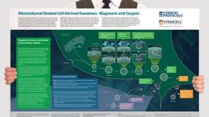 挂图Mesenchymal Stromal Cell-Derived Exosomes: Biogenesis and Cargoes Overview of exosome and microvesicle biogenesis pathways and potential therapeutic implications in the context of mesenchymal stromal cells


 沪公网安备31010102008431号