Interleukin-6 deficiency affects bone marrow stromal precursors, resulting in defective hematopoietic support.
Interleukin-6 (IL-6) is a critical factor in the regulation of stromal function and hematopoiesis. In vivo bromodeoxyuridine incorporation analysis indicates that the percentage of Lin(-)Sca-1(+) hematopoietic progenitors undergoing DNA synthesis is diminished in IL-6-deficient (IL-6(-/-)) bone marrow (BM) compared with wild-type BM. Reduced proliferation of IL-6(-/-) BM progenitors is also observed in IL-6(-/-) long-term BM cultures,which show defective hematopoietic support as measured by production of total cells,granulocyte macrophage-colony-forming units (CFU-GMs),and erythroid burst-forming units (BFU-Es). Seeding experiments of wild-type and IL-6(-/-) BM cells on irradiated wild-type or IL-6-deficient stroma indicate that the hematopoietic defect can be attributed to the stromal and not to the hematopoietic component. In IL-6(-/-) BM,stromal mesenchymal precursors,fibroblast CFUs (CFU-Fs),and stroma-initiating cells (SICs) are reduced to almost 50% of the wild-type BM value. Moreover,IL-6(-/-) stromata show increased CD34 and CD49e expression and reduced expression of the membrane antigens vascular cell adhesion molecule-1 (VCAM-1),Sca-1,CD49f,and Thy1. These data strongly suggest that IL-6 is an in vivo growth factor for mesenchymal precursors,which are in part implicated in the reduced longevity of the long-term repopulating stem cell compartment of IL-6(-/-) mice.
View Publication
产品号#:
03534
05501
05502
05350
28600
产品名:
MethoCult™ GF M3534
L-Calc™有限稀释软件
Eirew P et al. (DEC 2008)
Nature medicine 14 12 1384--9
A method for quantifying normal human mammary epithelial stem cells with in vivo regenerative ability.
Previous studies have demonstrated that normal mouse mammary tissue contains a rare subset of mammary stem cells. We now describe a method for detecting an analogous subpopulation in normal human mammary tissue. Dissociated cells are suspended with fibroblasts in collagen gels,which are then implanted under the kidney capsule of hormone-treated immunodeficient mice. After 2-8 weeks,the gels contain bilayered mammary epithelial structures,including luminal and myoepithelial cells,their in vitro clonogenic progenitors and cells that produce similar structures in secondary transplants. The regenerated clonogenic progenitors provide an objective indicator of input mammary stem cell activity and allow the frequency and phenotype of these human mammary stem cells to be determined by limiting-dilution analysis. This new assay procedure sets the stage for investigations of mechanisms regulating normal human mammary stem cells (and possibly stem cells in other tissues) and their relationship to human cancer stem cell populations.
View Publication
We studied the immunoregulatory features of murine mesenchymal stem cells (MSCs) in vitro and in vivo. MSCs inhibited T-cell receptor (TCR)-dependent and -independent proliferation but did not induce apoptosis on T cells. Such inhibition was paired with a decreased interferon (IFN)-gamma and tumor necrosis factor (TNF)-alpha production and was partially reversed by interleukin-2 (IL-2). Thus,we used MSCs to treat myelin oligodendrocyte glycoprotein (MOG)35-55-induced experimental autoimmune encephalomyelitis (EAE) in C57BL/6J mice. We injected intravenously 1 x 10(6) MSCs before disease onset (preventive protocol) and at different time points after disease occurrence (therapeutic protocol). MSC administration before disease onset strikingly ameliorated EAE. The therapeutic scheme was effective when MSCs were administered at disease onset and at the peak of disease but not after disease stabilization. Central nervous system (CNS) pathology showed decreased inflammatory infiltrates and demyelination in mice that received transplants of MSCs. T-cell response to MOG and mitogens from MSC-treated mice was inhibited and restored by IL-2 administration. Upon MSC transfection with the enhanced green fluorescent protein (eGFP),eGFP(+) cells were detected in the lymphoid organs of treated mice. These data suggest that the immunoregulatory properties of MSCs effectively interfere with the autoimmune attack in the course of EAE inducing an in vivo state of T-cell unresponsiveness occurring within secondary lymphoid organs.
View Publication
产品号#:
05501
05502
产品名:
Bieback K et al. (JAN 2004)
Stem cells (Dayton,Ohio) 22 4 625--34
Critical parameters for the isolation of mesenchymal stem cells from umbilical cord blood.
Evidence has emerged that mesenchymal stem cells (MSCs) represent a promising population for supporting new clinical concepts in cellular therapy. However,attempts to isolate MSCs from umbilical cord blood (UCB) of full-term deliveries have previously either failed or been characterized by a low yield. We investigated whether cells with MSC characteristics and multi-lineage differentiation potential can be cultivated from UCB of healthy newborns and whether yields might be maximized by optimal culture conditions or by defining UCB quality criteria. Using optimized isolation and culture conditions,in up to 63% of 59 low-volume UCB units,cells showing a characteristic mesenchymal morphology and immune phenotype (MSC-like cells) were isolated. These were similar to control MSCs from adult bone marrow (BM). The frequency of MSC-like cells ranged from 0 to 2.3 clones per 1 x 10(8) mononuclear cells (MNCs). The cell clones proliferated extensively with at least 20 population doublings within eight passages. In addition,osteogenic and chondrogenic differentiation demonstrated a multi-lineage capacity comparable with BM MSCs. However,in contrast to MSCs,MSC-like cells showed a reduced sensitivity to undergo adipogenic differentiation. Crucial points to isolate MSC-like cells from UCB were a time from collection to isolation of less than 15 hours,a net volume of more than 33 ml,and an MNC count of more than 1 x 10(8) MNCs. Because MSC-like cells can be isolated at high efficacy from full-term UCB donations,we regard UCB as an additional stem cell source for experimental and potentially clinical purposes.
View Publication

 EasySep™小鼠TIL(CD45)正选试剂盒


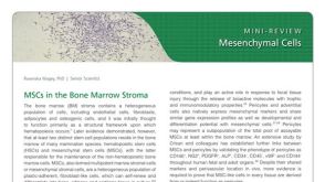 研究综述Mesenchymal Stromal Cells: Markers, Isolation and Culture, Differentiation, and Therapeutic Potential


 沪公网安备31010102008431号