Wu X et al. (APR 2011)
The Journal of biological chemistry 286 15 13512--21
p85alpha regulates osteoblast differentiation by cross-talking with the MAPK pathway.
Class IA phosphoinositide 3-kinase (PI3K) is involved in regulating many cellular functions including cell growth,proliferation,cell survival,and differentiation. The p85 regulatory subunit is a critical component of the PI3K signaling pathway. Mesenchymal stem cells (MSC) are multipotent cells that can be differentiated into osteoblasts (OBs),adipocytes,and chondrocytes under defined culture conditions. To determine whether p85α subunit of PI3K affects biological functions of MSCs,bone marrow-derived wild type (WT) and p85α-deficient (p85α(-/-)) cells were employed in this study. Increased cell growth,higher proliferation rate and reduced number of senescent cells were observed in MSCs lacking p85α compare with WT MSCs as evaluated by CFU-F assay,thymidine incorporation assay,and β-galactosidase staining,respectively. These functional changes are associated with the increased cell cycle,increased expression of cyclin D,cyclin E,and reduced expression of p16 and p19 in p85α(-/-) MSCs. In addition,a time-dependent reduction in alkaline phosphatase (ALP) activity and osteocalcin mRNA expression was observed in p85α(-/-) MSCs compared with WT MSCs,suggesting impaired osteoblast differentiation due to p85α deficiency in MSCs. The impaired p85α(-/-) osteoblast differentiation was associated with increased activation of Akt and MAPK. Importantly,bone morphogenic protein 2 (BMP2) was able to intensify the differentiation of osteoblasts derived from WT MSCs,whereas this process was significantly impaired as a result of p85α deficiency. Addition of LY294002,a PI3K inhibitor,did not alter the differentiation of osteoblasts in either genotype. However,application of PD98059,a Mek/MAPK inhibitor,significantly enhanced osteoblast differentiation in WT and p85α(-/-) MSCs. These results suggest that p85α plays an essential role in osteoblast differentiation from MSCs by repressing the activation of MAPK pathway.
View Publication
产品号#:
05501
05502
产品名:
Keller GM (DEC 1995)
Current opinion in cell biology 7 6 862--9
In vitro differentiation of embryonic stem cells.
Under appropriate conditions in culture,embryonic stem cells will differentiate and form embryoid bodies that have been shown to contain cells of the hematopoietic,endothelial,muscle and neuronal lineages. Many aspects of the lineage-specific differentiation programs observed within the embryoid bodies reflect those found in the embryo,indicating that this model system provides access to early cell populations that develop in a normal fashion. Recent studies involving the differentiation of genetically altered embryonic stem cells highlight the potential of this in vitro differentiation system for defining the function of genes in early development.
View Publication
产品号#:
06902
06952
00321
00322
00323
00324
00325
产品名:
Rubin MR et al. (JAN 2011)
The Journal of clinical endocrinology and metabolism 96 1 176--86
Parathyroid hormone stimulates circulating osteogenic cells in hypoparathyroidism.
CONTEXT: The osteoanabolic properties of PTH may be due to increases in the number and maturity of circulating osteogenic cells. Hypoparathyroidism is a useful clinical model because this hypothesis can be tested by administering PTH. OBJECTIVE: The objective of the study was to characterize circulating osteogenic cells in hypoparathyroid subjects during 12 months of PTH (1-84) administration. DESIGN: Osteogenic cells were characterized using flow cytometry and antibodies against osteocalcin,an osteoblast-specific protein product,and stem cell markers CD34 and CD146. Changes in bone formation from biochemical markers and quadruple-labeled transiliac crest bone biopsies (0 and 3 month time points) were correlated with measurements of circulating osteogenic cells. SETTING: The study was conducted at a clinical research center. PATIENTS: Nineteen control and 19 hypoparathyroid patients were included in the study. INTERVENTION: Intervention included the administration of PTH (1-84). RESULTS: Osteocalcin-positive cells were lower in hypoparathyroid subjects than controls (0.7 ± 0.1 vs. 2.0 ± 0.1%; P textless 0.0001),with greater coexpression of the early cell markers CD34 and CD146 among the osteocalcin-positive cells in the hypoparathyroid subjects (11.0 ± 1.0 vs. 5.6 ± 0.7%; P textless 0.001). With PTH (1-84) administration,the number of osteogenic cells increased 3-fold (P textless 0.0001),whereas the coexpression of the early cell markers CD34 and CD146 decreased. Increases in osteogenic cells correlated with circulating and histomorphometric indices of osteoblast function: N-terminal propeptide of type I procollagen (R(2) = 0.4,P ≤ 0.001),bone-specific alkaline phosphatase (R(2) = 0.3,P textless 0.001),osteocalcin (R(2) = 0.4,P textless 0.001),mineralized perimeter (R(2) = 0.5,P textless 0.001),mineral apposition rate (R(2) = 0.4,P = 0.003),and bone formation rate (R(2) = 0.5,P textless 0.001). CONCLUSIONS: It is likely that PTH stimulates bone formation by stimulating osteoblast development and maturation. Correlations between circulating osteogenic cells and histomorphometric indices of bone formation establish that osteoblast activity is being identified by this methodology.
View Publication
产品号#:
05404
产品名:
Rodrí et al. (MAY 2004)
Blood 103 9 3349--54
Interleukin-6 deficiency affects bone marrow stromal precursors, resulting in defective hematopoietic support.
Interleukin-6 (IL-6) is a critical factor in the regulation of stromal function and hematopoiesis. In vivo bromodeoxyuridine incorporation analysis indicates that the percentage of Lin(-)Sca-1(+) hematopoietic progenitors undergoing DNA synthesis is diminished in IL-6-deficient (IL-6(-/-)) bone marrow (BM) compared with wild-type BM. Reduced proliferation of IL-6(-/-) BM progenitors is also observed in IL-6(-/-) long-term BM cultures,which show defective hematopoietic support as measured by production of total cells,granulocyte macrophage-colony-forming units (CFU-GMs),and erythroid burst-forming units (BFU-Es). Seeding experiments of wild-type and IL-6(-/-) BM cells on irradiated wild-type or IL-6-deficient stroma indicate that the hematopoietic defect can be attributed to the stromal and not to the hematopoietic component. In IL-6(-/-) BM,stromal mesenchymal precursors,fibroblast CFUs (CFU-Fs),and stroma-initiating cells (SICs) are reduced to almost 50% of the wild-type BM value. Moreover,IL-6(-/-) stromata show increased CD34 and CD49e expression and reduced expression of the membrane antigens vascular cell adhesion molecule-1 (VCAM-1),Sca-1,CD49f,and Thy1. These data strongly suggest that IL-6 is an in vivo growth factor for mesenchymal precursors,which are in part implicated in the reduced longevity of the long-term repopulating stem cell compartment of IL-6(-/-) mice.
View Publication

 EasySep™小鼠TIL(CD45)正选试剂盒


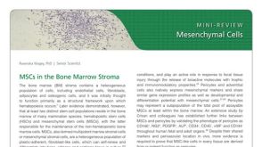 研究综述Mesenchymal Stromal Cells: Markers, Isolation and Culture, Differentiation, and Therapeutic Potential


 沪公网安备31010102008431号