Cunha B et al. (NOV 2015)
Journal of biotechnology 213 97--108
Exploring continuous and integrated strategies for the up- and downstream processing of human mesenchymal stem cells.
The integration of up- and downstream unit operations can result in the elimination of hold steps,thus decreasing the footprint,and ultimately can create robust closed system operations. This type of design is desirable for the bioprocess of human mesenchymal stem cells (hMSC),where high numbers of pure cells,at low volumes,need to be delivered for therapy applications. This study reports a proof of concept of the integration of a continuous perfusion culture in bioreactors with a tangential flow filtration (TFF) system for the concentration and washing of hMSC. Moreover,we have also explored a continuous alternative for concentrating hMSC. Results show that expanding cells in a continuous perfusion operation mode provided a higher expansion ratio,and led to a shift in cells' metabolism. TFF operated either in continuous or discontinuous allowed to concentrate cells,with high cell recovery (>80%) and viability (>95%); furthermore,continuous TFF permitted to operate longer with higher cell concentrations. Continuous diafiltration led to higher protein clearance (98%) with lower cell death,when comparing to discontinuous diafiltration. Overall,an integrated process allowed for a shorter process time,recovering 70% of viable hMSC (>95%),with no changes in terms of morphology,immunophenotype,proliferation capacity and multipotent differentiation potential.
View Publication
产品号#:
70022
70071
产品名:
Zhao Q et al. (JAN 2015)
Proceedings of the National Academy of Sciences of the United States of America 112 2 530--535
MSCs derived from iPSCs with a modified protocol are tumor-tropic but have much less potential to promote tumors than bone marrow MSCs.
Mesenchymal stem or stromal cells (MSCs) have many potential therapeutic applications including therapies for cancers and tissue damages caused by cancers or radical cancer treatments. However,tissue-derived MSCs such as bone marrow MSCs (BM-MSCs) may promote cancer progression and have considerable donor variations and limited expandability. These issues hinder the potential applications of MSCs,especially those in cancer patients. To circumvent these issues,we derived MSCs from transgene-free human induced pluripotent stem cells (iPSCs) efficiently with a modified protocol that eliminated the need of flow cytometric sorting. Our iPSC-derived MSCs were readily expandable,but still underwent senescence after prolonged culture and did not form teratomas. These iPSC-derived MSCs homed to cancers with efficiencies similar to BM-MSCs but were much less prone than BM-MSCs to promote the epithelial-mesenchymal transition,invasion,stemness,and growth of cancer cells. The observations were probably explained by the much lower expression of receptors for interleukin-1 and TGFβ,downstream protumor factors,and hyaluronan and its cofactor TSG6,which all contribute to the protumor effects of BM-MSCs. The data suggest that iPSC-derived MSCs prepared with the modified protocol are a safer and better alternative to BM-MSCs for therapeutic applications in cancer patients. The protocol is scalable and can be used to prepare the large number of cells required for off-the-shelf" therapies and bioengineering applications."
View Publication
产品号#:
01700
01705
05850
05857
05870
05875
85850
85857
85870
85875
01702
产品名:
ALDEFLUOR™ 试剂盒
ALDEFLUOR™ DEAB试剂, 1.5 mM, 1 mL
mTeSR™1
mTeSR™1
ALDEFLUOR™检测缓冲液
Gadkari R et al. (JUL 2014)
Regenerative medicine 9 4 453--465
Human embryonic stem cell derived-mesenchymal stem cells: an alternative mesenchymal stem cell source for regenerative medicine therapy.
AIM To enumerate and characterize mesenchymal stem cells (MSC) derived from human embryonic stem cells (hESC) for clinical application. MATERIALS & METHODS hESC were differentiated into hESC-MSC and characterized by the expression of surface markers using flow cytometry. hESC-MSC were evaluated with respect to growth kinetics,colony-forming potential,as well as osteogenic and adipogenic differentiation capacity. Immunosuppressive effects were assessed using peripheral blood mononuclear cell (PBMC) proliferation and cytotoxicity assays. RESULTS hESC-MSC showed similar morphology,and cell surface markers as adipose (AMSC) and bone marrow-derived MSC (BMSC). hESC-MSC exhibited a higher growth rate during early in vitro expansion and equivalent adipogenic and osteogenic differentiation and colony-forming potential as AMSC and BMSC. hESC-MSC demonstrated similar immunosuppressive effects as AMSC and BMSC. CONCLUSION hESC-MSC were comparable to BMSC and AMSC and hence can be used as an alternative source of MSC for clinical applications.
View Publication
Keller GM (DEC 1995)
Current opinion in cell biology 7 6 862--9
In vitro differentiation of embryonic stem cells.
Under appropriate conditions in culture,embryonic stem cells will differentiate and form embryoid bodies that have been shown to contain cells of the hematopoietic,endothelial,muscle and neuronal lineages. Many aspects of the lineage-specific differentiation programs observed within the embryoid bodies reflect those found in the embryo,indicating that this model system provides access to early cell populations that develop in a normal fashion. Recent studies involving the differentiation of genetically altered embryonic stem cells highlight the potential of this in vitro differentiation system for defining the function of genes in early development.
View Publication
产品号#:
06902
06952
00321
00322
00323
00324
00325
产品名:
Sessarego N et al. (MAR 2008)
Haematologica 93 3 339--46
Multipotent mesenchymal stromal cells from amniotic fluid: solid perspectives for clinical application.
BACKGROUND: Mesenchymal stromal cells are multipotent cells considered to be of great promise for use in regenerative medicine. However,the cell dose may be a critical factor in many clinical conditions and the yield resulting from the ex vivo expansion of mesenchymal stromal cells derived from bone marrow may be insufficient. Thus,alternative sources of mesenchymal stromal cells need to be explored. In this study,mesenchymal stromal cells were successfully isolated from second trimester amniotic fluid and analyzed for chromosomal stability to validate their safety for potential utilization as a cell therapy product. DESIGN AND METHODS: Mesenchymal stromal cells were expanded up to the sixth passage starting from amniotic fluid using different culture conditions to optimize large-scale production. RESULTS: The highest number of mesenchymal stromal cells derived from amniotic fluid was reached at a low plating density; in these conditions the expansion of mesenchymal stromal cells from amniotic fluid was significantly greater than that of adult bone marrow-derived mesenchymal stromal cells. Mesenchymal stromal cells from amniotic fluid represent a relatively homogeneous population of immature cells with immunosuppressive properties and extensive proliferative potential. Despite their high proliferative capacity in culture,we did not observe any karyotypic abnormalities or transformation potential in vitro nor any tumorigenic effect in vivo. CONCLUSIONS: Fetal mesenchymal stromal cells can be extensively expanded from amniotic fluid,showing no karyotypic abnormalities or transformation potential in vitro and no tumorigenic effect in vivo. They represent a relatively homogeneous population of immature mesenchymal stromal cells with long telomeres,immunosuppressive properties and extensive proliferative potential. Our results indicate that amniotic fluid represents a rich source of mesenchymal stromal cells suitable for banking to be used when large amounts of cells are required.
View Publication
Wu X et al. (APR 2011)
The Journal of biological chemistry 286 15 13512--21
p85alpha regulates osteoblast differentiation by cross-talking with the MAPK pathway.
Class IA phosphoinositide 3-kinase (PI3K) is involved in regulating many cellular functions including cell growth,proliferation,cell survival,and differentiation. The p85 regulatory subunit is a critical component of the PI3K signaling pathway. Mesenchymal stem cells (MSC) are multipotent cells that can be differentiated into osteoblasts (OBs),adipocytes,and chondrocytes under defined culture conditions. To determine whether p85α subunit of PI3K affects biological functions of MSCs,bone marrow-derived wild type (WT) and p85α-deficient (p85α(-/-)) cells were employed in this study. Increased cell growth,higher proliferation rate and reduced number of senescent cells were observed in MSCs lacking p85α compare with WT MSCs as evaluated by CFU-F assay,thymidine incorporation assay,and β-galactosidase staining,respectively. These functional changes are associated with the increased cell cycle,increased expression of cyclin D,cyclin E,and reduced expression of p16 and p19 in p85α(-/-) MSCs. In addition,a time-dependent reduction in alkaline phosphatase (ALP) activity and osteocalcin mRNA expression was observed in p85α(-/-) MSCs compared with WT MSCs,suggesting impaired osteoblast differentiation due to p85α deficiency in MSCs. The impaired p85α(-/-) osteoblast differentiation was associated with increased activation of Akt and MAPK. Importantly,bone morphogenic protein 2 (BMP2) was able to intensify the differentiation of osteoblasts derived from WT MSCs,whereas this process was significantly impaired as a result of p85α deficiency. Addition of LY294002,a PI3K inhibitor,did not alter the differentiation of osteoblasts in either genotype. However,application of PD98059,a Mek/MAPK inhibitor,significantly enhanced osteoblast differentiation in WT and p85α(-/-) MSCs. These results suggest that p85α plays an essential role in osteoblast differentiation from MSCs by repressing the activation of MAPK pathway.
View Publication
产品号#:
05501
05502
产品名:
Rubin MR et al. (JAN 2011)
The Journal of clinical endocrinology and metabolism 96 1 176--86
Parathyroid hormone stimulates circulating osteogenic cells in hypoparathyroidism.
CONTEXT: The osteoanabolic properties of PTH may be due to increases in the number and maturity of circulating osteogenic cells. Hypoparathyroidism is a useful clinical model because this hypothesis can be tested by administering PTH. OBJECTIVE: The objective of the study was to characterize circulating osteogenic cells in hypoparathyroid subjects during 12 months of PTH (1-84) administration. DESIGN: Osteogenic cells were characterized using flow cytometry and antibodies against osteocalcin,an osteoblast-specific protein product,and stem cell markers CD34 and CD146. Changes in bone formation from biochemical markers and quadruple-labeled transiliac crest bone biopsies (0 and 3 month time points) were correlated with measurements of circulating osteogenic cells. SETTING: The study was conducted at a clinical research center. PATIENTS: Nineteen control and 19 hypoparathyroid patients were included in the study. INTERVENTION: Intervention included the administration of PTH (1-84). RESULTS: Osteocalcin-positive cells were lower in hypoparathyroid subjects than controls (0.7 ± 0.1 vs. 2.0 ± 0.1%; P textless 0.0001),with greater coexpression of the early cell markers CD34 and CD146 among the osteocalcin-positive cells in the hypoparathyroid subjects (11.0 ± 1.0 vs. 5.6 ± 0.7%; P textless 0.001). With PTH (1-84) administration,the number of osteogenic cells increased 3-fold (P textless 0.0001),whereas the coexpression of the early cell markers CD34 and CD146 decreased. Increases in osteogenic cells correlated with circulating and histomorphometric indices of osteoblast function: N-terminal propeptide of type I procollagen (R(2) = 0.4,P ≤ 0.001),bone-specific alkaline phosphatase (R(2) = 0.3,P textless 0.001),osteocalcin (R(2) = 0.4,P textless 0.001),mineralized perimeter (R(2) = 0.5,P textless 0.001),mineral apposition rate (R(2) = 0.4,P = 0.003),and bone formation rate (R(2) = 0.5,P textless 0.001). CONCLUSIONS: It is likely that PTH stimulates bone formation by stimulating osteoblast development and maturation. Correlations between circulating osteogenic cells and histomorphometric indices of bone formation establish that osteoblast activity is being identified by this methodology.
View Publication

 EasySep™小鼠TIL(CD45)正选试剂盒


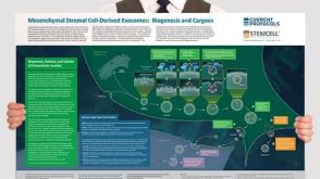 挂图Mesenchymal Stromal Cell-Derived Exosomes: Biogenesis and Cargoes Overview of exosome and microvesicle biogenesis pathways and potential therapeutic implications in the context of mesenchymal stromal cells


 沪公网安备31010102008431号