Russo FP et al. (MAY 2006)
Gastroenterology 130 6 1807--21
The bone marrow functionally contributes to liver fibrosis.
BACKGROUND & AIMS: Bone marrow (BM) cells may transdifferentiate into or fuse with organ parenchymal cells. BM therapy shows promise in murine models of cirrhosis,and clinical trials of bone marrow stem cell therapy for organ healing are underway. However,the BM may contribute to scar-forming myofibroblasts in various organs including the liver. We have studied this axis of regeneration and scarring in murine models of cirrhosis,including an assessment of the temporal and functional contribution of the BM-derived myofibroblasts. METHODS: Female mice were lethally irradiated and received male BM transplants. Carbon tetrachloride or thioacetamide was used to induce cirrhosis. BM-derived cells were tracked through in situ hybridization for the Y chromosome. BM transplants from 2 strains of transgenic mice were used to detect intrahepatic collagen production. RESULTS: In the cirrhotic liver,the contribution of BM to parenchymal regeneration was minor (0.6%); by contrast,the BM contributed significantly to hepatic stellate cell (68%) and myofibroblast (70%) populations. These BM-derived cells were found to be active for collagen type 1 transcription in 2 independent assays and could influence the fibrotic response to organ injury. These BM-derived myofibroblasts did not occur through cell fusion between BM-derived cells and indigenous hepatic cells but,instead,originated largely from the BM's mesenchymal stem cells. CONCLUSIONS: The BM contributes functionally and significantly to liver fibrosis and is a potential therapeutic target in liver fibrosis. Clinical trials of BM cell therapy for liver regeneration should be vigilant for the possibility of enhanced organ fibrosis.
View Publication
Mesenchymal stem cells can be differentiated into endothelial cells in vitro.
Human bone marrow-derived mesenchymal stem cells (MSCs) have the potential to differentiate into mesenchymal tissues like osteocytes,chondrocytes,and adipocytes in vivo and in vitro. The aim of this study was to investigate the in vitro differentiation of MSCs into cells of the endothelial lineage. MSCs were generated out of mononuclear bone marrow cells from healthy donors separated by density gradient centrifugation. Cells were characterized by flow cytometry using a panel of monoclonal antibodies and were tested for their potential to differentiate along different mesenchymal lineages. Isolated MSCs were positive for the markers CD105,CD73,CD166,CD90,and CD44 and negative for typical hematopoietic and endothelial markers. They were able to differentiate into adipocytes and osteocytes after cultivation in respective media. Differentiation into endothelial-like cells was induced by cultivation of confluent cells in the presence of 2% fetal calf serum and 50 ng/ml vascular endothelial growth factor. Laser scanning cytometry analysis of the confluent cells in situ showed a strong increase of expression of endothelial-specific markers like KDR and FLT-1,and immunofluorescence analysis showed typical expression of the von Willebrand factor. The functional behavior of the differentiated cells was tested with an in vitro angiogenesis test kit where cells formed characteristic capillary-like structures. We could show the differentiation of expanded adult human MSCs into cells with phenotypic and functional features of endothelial cells. These predifferentiated cells provide new options for engineering of artificial tissues based on autologous MSCs and vascularized engineered tissues.
View Publication
产品号#:
05401
产品名:
MesenCult™ MSC 基础培养基(人)
Vaysse L et al. (FEB 2004)
The Journal of biological chemistry 279 7 5555--64
Development of a self-assembling nuclear targeting vector system based on the tetracycline repressor protein.
The ultimate destination for most gene therapy vectors is the nucleus and nuclear import of potentially therapeutic DNA is one of the major barriers for nonviral vectors. We have developed a novel approach of attaching a nuclear localization sequence (NLS) peptide to DNA in a non-essential position,by generating a fusion between the tetracycline repressor protein TetR and the SV40-derived NLS peptide. The high affinity and specificity of TetR for the short DNA sequence tetO was used in these studies to bind the NLS to DNA as demonstrated by the reduced electrophoretic mobility of the TetR.tetO-DNA complexes. The protein TetR-NLS,but not control protein TetR,specifically enhances gene expression from lipofected tetO-containing DNA between 4- and 16-fold. The specific enhancement is observed in a variety of cell types,including primary and growth-arrested cells. Intracellular trafficking studies demonstrate an increased accumulation of fluorescence labeled DNA in the nucleus after TetR-NLS binding. In comparison,binding studies using the similar fusion of peptide nucleic acid (PNA) with NLS peptide,demonstrate specific binding of PNA to plasmid DNA. However,although we observed a 2-8.5-fold increase in plasmid-mediated luciferase activity with bis-PNA-NLS,control bis-PNA without an NLS sequence gave a similar increase,suggesting that the effect may not be because of a specific bis-PNA-NLS-mediated enhancement of nuclear transfer of the plasmid. Overall,we found TetRNLS-enhanced plasmid-mediated transgene expression at a similar level to that by bis-PNA-NLS or bis-PNA alone but specific to nuclear uptake and significantly more reliable and reproducible.
View Publication
产品号#:
05401
05402
05411
产品名:
MesenCult™ MSC 基础培养基(人)
MesenCult™ MSC 刺激补充剂(人)
MesenCult™ 增殖试剂盒(人)
Lee OK et al. (MAR 2004)
Blood 103 5 1669--75
Isolation of multipotent mesenchymal stem cells from umbilical cord blood.
It is well accepted that umbilical cord blood has been a source for hematopoietic stem cells. However,controversy exists as to whether cord blood can serve as a source of mesenchymal stem cells,which can differentiate into cells of different connective tissue lineages such as bone,cartilage,and fat,and little success has been reported in the literature about the isolation of such cells from cord blood. Here we report a novel method to obtain single cell-derived,clonally expanded mesenchymal stem cells that are of multilineage differentiation potential by negative immunoselection and limiting dilution. The immunophenotype of these clonally expanded cells is consistent with that reported for bone marrow mesenchymal stem cells. Under appropriate induction conditions,these cells can differentiate into bone,cartilage,and fat. Surprisingly,these cells were also able to differentiate into neuroglial- and hepatocyte-like cells under appropriate induction conditions and,thus,these cells may be more than mesenchymal stem cells as evidenced by their ability to differentiate into cell types of all 3 germ layers. In conclusion,umbilical cord blood does contain mesenchymal stem cells and should not be regarded as medical waste. It can serve as an alternative source of mesenchymal stem cells to bone marrow.
View Publication
产品号#:
15128
15168
产品名:
RosetteSep™人间充质干细胞富集抗体混合物
RosetteSep™人间充质干细胞富集抗体混合物
Keller GM (DEC 1995)
Current opinion in cell biology 7 6 862--9
In vitro differentiation of embryonic stem cells.
Under appropriate conditions in culture,embryonic stem cells will differentiate and form embryoid bodies that have been shown to contain cells of the hematopoietic,endothelial,muscle and neuronal lineages. Many aspects of the lineage-specific differentiation programs observed within the embryoid bodies reflect those found in the embryo,indicating that this model system provides access to early cell populations that develop in a normal fashion. Recent studies involving the differentiation of genetically altered embryonic stem cells highlight the potential of this in vitro differentiation system for defining the function of genes in early development.
View Publication
产品号#:
06902
06952
00321
00322
00323
00324
00325
产品名:
Sessarego N et al. (MAR 2008)
Haematologica 93 3 339--46
Multipotent mesenchymal stromal cells from amniotic fluid: solid perspectives for clinical application.
BACKGROUND: Mesenchymal stromal cells are multipotent cells considered to be of great promise for use in regenerative medicine. However,the cell dose may be a critical factor in many clinical conditions and the yield resulting from the ex vivo expansion of mesenchymal stromal cells derived from bone marrow may be insufficient. Thus,alternative sources of mesenchymal stromal cells need to be explored. In this study,mesenchymal stromal cells were successfully isolated from second trimester amniotic fluid and analyzed for chromosomal stability to validate their safety for potential utilization as a cell therapy product. DESIGN AND METHODS: Mesenchymal stromal cells were expanded up to the sixth passage starting from amniotic fluid using different culture conditions to optimize large-scale production. RESULTS: The highest number of mesenchymal stromal cells derived from amniotic fluid was reached at a low plating density; in these conditions the expansion of mesenchymal stromal cells from amniotic fluid was significantly greater than that of adult bone marrow-derived mesenchymal stromal cells. Mesenchymal stromal cells from amniotic fluid represent a relatively homogeneous population of immature cells with immunosuppressive properties and extensive proliferative potential. Despite their high proliferative capacity in culture,we did not observe any karyotypic abnormalities or transformation potential in vitro nor any tumorigenic effect in vivo. CONCLUSIONS: Fetal mesenchymal stromal cells can be extensively expanded from amniotic fluid,showing no karyotypic abnormalities or transformation potential in vitro and no tumorigenic effect in vivo. They represent a relatively homogeneous population of immature mesenchymal stromal cells with long telomeres,immunosuppressive properties and extensive proliferative potential. Our results indicate that amniotic fluid represents a rich source of mesenchymal stromal cells suitable for banking to be used when large amounts of cells are required.
View Publication
Immunophenotype of human adipose-derived cells: temporal changes in stromal-associated and stem cell-associated markers.
Adipose tissue represents an abundant and accessible source of multipotent adult stem cells and is used by many investigators for tissue engineering applications; however,not all laboratories use cells at equivalent stages of isolation and passage. We have compared the immunophenotype of freshly isolated human adipose tissue-derived stromal vascular fraction (SVF) cells relative to serial-passaged adipose-derived stem cells (ASCs). The initial SVF cells contained colony-forming unit fibroblasts at a frequency of 1:32. Colony-forming unit adipocytes and osteoblasts were present in the SVF cells at comparable frequencies (1:28 and 1:16,respectively). The immunophenotype of the adipose-derived cells based on flow cytometry changed progressively with adherence and passage. Stromal cell-associated markers (CD13,CD29,CD44,CD63,CD73,CD90,CD166) were initially low on SVF cells and increased significantly with successive passages. The stem cell-associated marker CD34 was at peak levels in the SVF cells and/or early-passage ASCs and remained present,although at reduced levels,throughout the culture period. Aldehyde dehydrogenase and the multidrug-resistance transport protein (ABCG2),both of which have been used to identify and characterize hematopoietic stem cells,are expressed by SVF cells and ASCs at detectable levels. Endothelial cell-associated markers (CD31,CD144 or VE-cadherin,vascular endothelial growth factor receptor 2,von Willebrand factor) were expressed on SVF cells and did not change significantly with serial passage. Thus,the adherence to plastic and subsequent expansion of human adipose-derived cells in fetal bovine serum-supplemented medium selects for a relatively homogeneous cell population,enriching for cells expressing a stromal immunophenotype,compared with the heterogeneity of the crude SVF.
View Publication
产品号#:
01700
01705
01702
产品名:
ALDEFLUOR™ 试剂盒
ALDEFLUOR™ DEAB试剂, 1.5 mM, 1 mL
ALDEFLUOR™检测缓冲液
Rubin MR et al. (JAN 2011)
The Journal of clinical endocrinology and metabolism 96 1 176--86
Parathyroid hormone stimulates circulating osteogenic cells in hypoparathyroidism.
CONTEXT: The osteoanabolic properties of PTH may be due to increases in the number and maturity of circulating osteogenic cells. Hypoparathyroidism is a useful clinical model because this hypothesis can be tested by administering PTH. OBJECTIVE: The objective of the study was to characterize circulating osteogenic cells in hypoparathyroid subjects during 12 months of PTH (1-84) administration. DESIGN: Osteogenic cells were characterized using flow cytometry and antibodies against osteocalcin,an osteoblast-specific protein product,and stem cell markers CD34 and CD146. Changes in bone formation from biochemical markers and quadruple-labeled transiliac crest bone biopsies (0 and 3 month time points) were correlated with measurements of circulating osteogenic cells. SETTING: The study was conducted at a clinical research center. PATIENTS: Nineteen control and 19 hypoparathyroid patients were included in the study. INTERVENTION: Intervention included the administration of PTH (1-84). RESULTS: Osteocalcin-positive cells were lower in hypoparathyroid subjects than controls (0.7 ± 0.1 vs. 2.0 ± 0.1%; P textless 0.0001),with greater coexpression of the early cell markers CD34 and CD146 among the osteocalcin-positive cells in the hypoparathyroid subjects (11.0 ± 1.0 vs. 5.6 ± 0.7%; P textless 0.001). With PTH (1-84) administration,the number of osteogenic cells increased 3-fold (P textless 0.0001),whereas the coexpression of the early cell markers CD34 and CD146 decreased. Increases in osteogenic cells correlated with circulating and histomorphometric indices of osteoblast function: N-terminal propeptide of type I procollagen (R(2) = 0.4,P ≤ 0.001),bone-specific alkaline phosphatase (R(2) = 0.3,P textless 0.001),osteocalcin (R(2) = 0.4,P textless 0.001),mineralized perimeter (R(2) = 0.5,P textless 0.001),mineral apposition rate (R(2) = 0.4,P = 0.003),and bone formation rate (R(2) = 0.5,P textless 0.001). CONCLUSIONS: It is likely that PTH stimulates bone formation by stimulating osteoblast development and maturation. Correlations between circulating osteogenic cells and histomorphometric indices of bone formation establish that osteoblast activity is being identified by this methodology.
View Publication

 EasySep™小鼠TIL(CD45)正选试剂盒


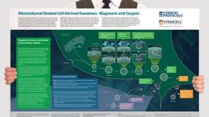 挂图Mesenchymal Stromal Cell-Derived Exosomes: Biogenesis and Cargoes Overview of exosome and microvesicle biogenesis pathways and potential therapeutic implications in the context of mesenchymal stromal cells


 沪公网安备31010102008431号