
 EasySep™小鼠TIL(CD45)正选试剂盒


技术资料
-
-
-
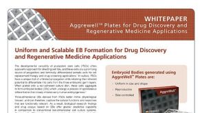 技术公告Uniform and Scalable EB Formation for Drug Discovery and Regenerative Medicine Applications
细胞类型:
PSC衍生造血细胞,多能干细胞,PSC衍生内胚层,PSC衍生心肌细胞 ,PSC衍生神经细胞
发布日期: 06/01/2012 -
-


 沪公网安备31010102008431号