Reprogramming Malignant Cancer Cells toward a Benign Phenotype following Exposure to Human Embryonic Stem Cell Microenvironment.
The embryonic microenvironment is well known to be non-permissive for tumor development because early developmental signals naturally suppress the expression of proto-oncogenes. In an analogous manner,mimicking an early embryonic environment during embryonic stem cell culture has been shown to suppress oncogenic phenotypes of cancer cells. Exosomes derived from human embryonic stem cells harbor substances that mirror the content of the cells of origin and have been reported to reprogram hematopoietic stem/progenitor cells via horizontal transfer of mRNA and proteins. However,the possibility that these embryonic stem cells-derived exosomes might be the main effectors of the anti-tumor effect mediated by the embryonic stem cells has not been explored yet. The present study aims to investigate whether exosomes derived from human embryonic stem cells can reprogram malignant cancer cells to a benign stage and reduce their tumorigenicity. We show that the embryonic stem cell-conditioned medium contains factors that inhibit cancer cell growth and tumorigenicity in vitro and in vivo. Moreover,we demonstrate that exosomes derived from human embryonic stem cells display anti-proliferation and pro-apoptotic effects,and decrease tumor size in a xenograft model. These exosomes are also able to transfer their cargo into target cancer cells,inducing a dose-dependent increase in SOX2,OCT4 and Nanog proteins,leading to a dose-dependent decrease of cancer cell growth and tumorigenicity. This study shows for the first time that human embryonic stem cell-derived exosomes play an important role in the tumor suppressive activity displayed by human embryonic stem cells.
View Publication
产品号#:
85850
85857
85870
85875
产品名:
mTeSR™1
mTeSR™1
Bershteyn M et al. (APR 2017)
Cell stem cell 20 4 435--449.e4
Human iPSC-Derived Cerebral Organoids Model Cellular Features of Lissencephaly and Reveal Prolonged Mitosis of Outer Radial Glia.
Classical lissencephaly is a genetic neurological disorder associated with mental retardation and intractable epilepsy,and Miller-Dieker syndrome (MDS) is the most severe form of the disease. In this study,to investigate the effects of MDS on human progenitor subtypes that control neuronal output and influence brain topology,we analyzed cerebral organoids derived from control and MDS-induced pluripotent stem cells (iPSCs) using time-lapse imaging,immunostaining,and single-cell RNA sequencing. We saw a cell migration defect that was rescued when we corrected the MDS causative chromosomal deletion and severe apoptosis of the founder neuroepithelial stem cells,accompanied by increased horizontal cell divisions. We also identified a mitotic defect in outer radial glia,a progenitor subtype that is largely absent from lissencephalic rodents but critical for human neocortical expansion. Our study,therefore,deepens our understanding of MDS cellular pathogenesis and highlights the broad utility of cerebral organoids for modeling human neurodevelopmental disorders.
View Publication
Human Induced Pluripotent Stem Cell-Derived Macrophages Share Ontogeny with MYB-Independent Tissue-Resident Macrophages.
Tissue-resident macrophages,such as microglia,Kupffer cells,and Langerhans cells,derive from Myb-independent yolk sac (YS) progenitors generated before the emergence of hematopoietic stem cells (HSCs). Myb-independent YS-derived resident macrophages self-renew locally,independently of circulating monocytes and HSCs. In contrast,adult blood monocytes,as well as infiltrating,gut,and dermal macrophages,derive from Myb-dependent HSCs. These findings are derived from the mouse,using gene knockouts and lineage tracing,but their applicability to human development has not been formally demonstrated. Here,we use human induced pluripotent stem cells (iPSCs) as a tool to model human hematopoietic development. By using a CRISPR-Cas9 knockout strategy,we show that human iPSC-derived monocytes/macrophages develop in an MYB-independent,RUNX1-,and SPI1 (PU.1)-dependent fashion. This result makes human iPSC-derived macrophages developmentally related to and a good model for MYB-independent tissue-resident macrophages,such as alveolar and kidney macrophages,microglia,Kupffer cells,and Langerhans cells.
View Publication
产品号#:
85850
85857
85870
85875
产品名:
mTeSR™1
mTeSR™1
Chou S-J et al. (APR 2017)
International journal of cardiology 232 255--263
Energy utilization of induced pluripotent stem cell-derived cardiomyocyte in Fabry disease.
BACKGROUND Fabry disease (FD) is a lysosomal storage disease in which glycosphingolipids (GB3) accumulate in organs of the human body,leading to idiopathic hypertrophic cardiomyopathy and target organ damage. Its pathophysiology is still poorly understood. OBJECTIVES We aimed to generate patient-specific induced pluripotent stem cells (iPSC) from FD patients presenting cardiomyopathy to determine whether the model could recapitulate key features of the disease phenotype and to investigate the energy metabolism in Fabry disease. METHODS Peripheral blood mononuclear cells from a 30-year-old Chinese man with a diagnosis of Fabry disease,GLA gene (IVS4+919G>A) mutation were reprogrammed into iPSCs and differentiated into iPSC-CMs and energy metabolism was analyzed in iPSC-CMs. RESULTS The FD-iPSC-CMs recapitulated numerous aspects of the FD phenotype including reduced GLA activity,cellular hypertrophy,GB3 accumulation and impaired contractility. Decreased energy metabolism with energy utilization shift to glycolysis was observed,but the decreased energy metabolism was not modified by enzyme rescue replacement (ERT) in FD-iPSCs-CMs. CONCLUSION This model provided a promising in vitro model for the investigation of the underlying disease mechanism and development of novel therapeutic strategies for FD. This potential remedy for enhancing the energetic network and utility efficiency warrants further study to identify novel therapies for the disease.
View Publication
产品号#:
85850
85857
85870
85875
产品名:
mTeSR™1
mTeSR™1
Gupta S et al. (DEC 2017)
Journal of Neurochemistry
Fibroblast growth factor 2 regulates activity and gene expression of human post-mitotic excitatory neurons
Many neuropsychiatric disorders are thought to result from subtle changes in neural circuit formation. We used human embryonic stem cells and induced pluripotent stem cells (hiPSCs) to model mature,post-mitotic excitatory neurons and examine effects of fibroblast growth factor 2 (FGF2). FGF2 gene expression is known to be altered in brain regions of major depressive disorder (MDD) patients and FGF2 has anti-depressive effects in animal models of depression. We generated stable inducible neurons (siNeurons) conditionally expressing human neurogenin-2 (NEUROG2) to generate a homogenous population of post-mitotic excitatory neurons and study the functional as well as the transcriptional effects of FGF2. Upon induction of NEUROG2 with doxycycline,the vast majority of cells are post-mitotic,and the gene expression profile recapitulates that of excitatory neurons within 6 days. Using hES cell lines that inducibly express NEUROG2 as well as GCaMP6f,we were able to characterize spontaneous calcium activity in these neurons and show that calcium transients increase in the presence of FGF2. The FGF2-responsive genes were determined by RNA-Seq. FGF2-regulated genes previously identified in non-neuronal cell types were up-regulated (EGR1,ETV4,SPRY4,and DUSP6) as a result of chronic FGF2 treatment of siNeurons. Novel neuron-specific genes were also identified that may mediate FGF2-dependent increases in synaptic efficacy including NRXN3,SYT2,and GALR1. Since several of these genes have been implicated in MDD previously,these results will provide the basis for more mechanistic studies of the role of FGF2 in MDD.
View Publication
K. B. Langer et al. (APR 2018)
Stem cell reports 10 4 1282--1293
Retinal Ganglion Cell Diversity and Subtype Specification from Human Pluripotent Stem Cells.
Retinal ganglion cells (RGCs) are the projection neurons of the retina and transmit visual information to postsynaptic targets in the brain. While this function is shared among nearly all RGCs,this class of cell is remarkably diverse,comprised of multiple subtypes. Previous efforts have identified numerous RGC subtypes in animal models,but less attention has been paid to human RGCs. Thus,efforts of this study examined the diversity of RGCs differentiated from human pluripotent stem cells (hPSCs) and characterized defined subtypes through the expression of subtype-specific markers. Further investigation of these subtypes was achieved using single-cell transcriptomics,confirming the combinatorial expression of molecular markers associated with these subtypes,and also provided insight into more subtype-specific markers. Thus,the results of this study describe the derivation of RGC subtypes from hPSCs and will support the future exploration of phenotypic and functional diversity within human RGCs.
View Publication
Alkaline phosphatase-positive colony formation is a sensitive, specific, and quantitative indicator of undifferentiated human embryonic stem cells.
Human embryonic stem cells (hESCs) can be maintained in vitro as immortal pluripotent cells but remain responsive to many differentiation-inducing signals. Investigation of the initial critical events involved in differentiation induction would be greatly facilitated if a specific,robust,and quantitative assay for pluripotent hESCs with self-renewal potential were available. Here we describe the results of a series of experiments to determine whether the formation of adherent alkaline phosphatase-positive (AP(+)) colonies under conditions optimized for propagating undifferentiated hESCs would meet this need. The findings can be summarized as follows. (a) Most colonies obtained under these conditions consist of textgreateror=30 AP(+) cells that coexpress OCT4,NANOG,SSEA3,SSEA4,TRA-1-60,and TRA-1-81. (b) Most such colonies are derived from SSEA3(+) cells. (c) Primary colonies contain cells that produce secondary colonies of the same composition,including cells that initiate multilineage differentiation in embryoid bodies (EBs). (d) Colony formation is independent of plating density or the colony-forming cell (CFC) content of the test population over a wide range of cell concentrations. (e) CFC frequencies decrease when differentiation is induced by exposure either to retinoic acid or to conditions that stimulate EB formation. Interestingly,this loss of AP(+) clonogenic potential also occurs more rapidly than the loss of SSEA3 or OCT4 expression. The CFC assay thus provides a simple,reliable,broadly applicable,and highly specific functional assay for quantifying undifferentiated hESCs with self-renewal potential. Its use under standardized assay conditions should enhance future elucidation of the mechanisms that regulate hESC propagation and their early differentiation.
View Publication


 EasySep™小鼠TIL(CD45)正选试剂盒




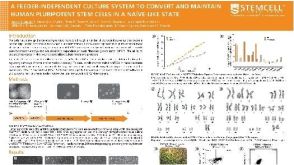 科学海报A Feeder-Independent Culture System to Convert and Maintain Human Pluripotent Stem Cells in a Naïve-Like State
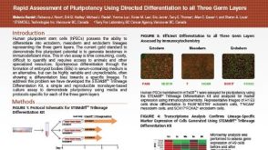 科学海报Rapid Assessment of Pluripotency Using Directed Differentiation to all Three Germ Layers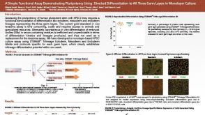

 沪公网安备31010102008431号