Bhinge A et al. (JUN 2014)
EMBO Journal 33 11 1271--1283
MiR-135b is a direct PAX6 target and specifies human neuroectoderm by inhibiting TGF-$\$/BMP signaling.
Several transcription factors (TFs) have been implicated in neuroectoderm (NE) development,and recently,the TF PAX6 was shown to be critical for human NE specification. However,microRNA networks regulating human NE development have been poorly documented. We hypothesized that microRNAs activated by PAX6 should promote NE development. Using a genomics approach,we identified PAX6 binding sites and active enhancers genome-wide in an in vitro model of human NE development that was based on neural differentiation of human embryonic stem cells (hESC). PAX6 binding to active enhancers was found in the proximity of several microRNAs,including hsa-miR-135b. MiR-135b was activated during NE development,and ectopic expression of miR-135b in hESC promoted differentiation toward NE. MiR-135b promotes neural conversion by targeting components of the TGF-β and BMP signaling pathways,thereby inhibiting differentiation into alternate developmental lineages. Our results demonstrate a novel TF-miRNA module that is activated during human neuroectoderm development and promotes the irreversible fate specification of human pluripotent cells toward the neural lineage.
View Publication
产品号#:
05850
05857
05870
05875
85850
85857
85870
85875
产品名:
mTeSR™1
mTeSR™1
Chin ACP et al. (JUN 2010)
Stem cells and development 19 6 753--61
Defined and serum-free media support undifferentiated human embryonic stem cell growth.
Four commercially available serum-free and defined culture media tested on 2 human embryonic stem cell (hESC) lines were all found to support undifferentiated growth for textgreater10 continuous passages. For hESC cultured with defined StemPro and mTeSR1 media,the cells were maintained feeder-free on culture dishes coated with extracellular matrices (ECMs) with no requirement of feeder-conditioned media (CM). For xeno-free serum replacer (XSR),HEScGRO,and KnockOut media,mitotically inactivated human foreskin feeders (hFFs) were required for hESC growth. Under the different media conditions,cells continued to exhibit alkaline phosphatase activity and expressed undifferentiated hESC markers Oct-4,stage-specific embryonic antigens 4 (SSEA-4),and Tra-1-60. In addition,hESC maintained the expression of podocalyxin-like protein-1 (PODXL),an antigen recently reported in another study to be present in undifferentiated hESC. The cytotoxic antibody mAb 84 binds via PODXL expressed on hESC surface and kills textgreater90% of hESC within 45 min of incubation. When these cells were spontaneously differentiated to form embryoid bodies,derivatives representing the 3 germ layers were obtained. Injection of hESC into animal models resulted in teratomas and the formation of tissue types indicative of ectodermal,endodermal,and mesodermal lineages were observed. Our data also suggested that StemPro and mTeSR1 media were more optimal for hESC proliferation compared to cells grown on CM because the growth rate of hESC increased by 30%-40%,higher split ratio was thus required for weekly passaging. This is advantageous for the large-scale cultivation of hESC required in clinical applications.
View Publication
产品号#:
05850
05857
05870
05875
85850
85857
85870
85875
产品名:
mTeSR™1
mTeSR™1
Tan H-K et al. (MAY 2014)
Stem cells translational medicine 3 5 586--98
Human finger-prick induced pluripotent stem cells facilitate the development of stem cell banking.
Induced pluripotent stem cells (iPSCs) derived from somatic cells of patients can be a good model for studying human diseases and for future therapeutic regenerative medicine. Current initiatives to establish human iPSC (hiPSC) banking face challenges in recruiting large numbers of donors with diverse diseased,genetic,and phenotypic representations. In this study,we describe the efficient derivation of transgene-free hiPSCs from human finger-prick blood. Finger-prick sample collection can be performed on a do-it-yourself" basis by donors and sent to the hiPSC facility for reprogramming. We show that single-drop volumes of finger-prick samples are sufficient for performing cellular reprogramming�
View Publication
产品号#:
05850
05857
05870
05875
09600
09650
85850
85857
85870
85875
产品名:
StemSpan™ SFEM
StemSpan™ SFEM
mTeSR™1
mTeSR™1
Hughes JN et al. (MAR 2014)
Differentiation; research in biological diversity 87 3-4 101--110
Regulation of pluripotent cell differentiation by a small molecule, staurosporine
Research in the embryo and in culture has resulted in a sophisticated understanding of many regulators of pluripotent cell differentiation. As a consequence,protocols for the differentiation of pluripotent cells generally rely on a combination of exogenous growth factors and endogenous signalling. Little consideration has been given to manipulating other pathways to achieve pluripotent cell differentiation. The integrity of cell:cell contacts has been shown to influence lineage choice during pluripotent cell differentiation,with disruption of cell:cell contacts promoting mesendoderm formation and maintenance of cell:cell contacts resulting in the preferential formation of neurectoderm. Staurosporine is a broad spectrum inhibitor of serine/threonine kinases which has several effects on cell function,including interruption of cell:cell contacts,decreasing focal contact size,inducing epithelial to mesenchyme transition (EMT) and promoting cell differentiation. The possibility that staurosporine could influence lineage choice from pluripotent cells in culture was investigated. The addition of staurosporine to differentiating mouse EPL resulted in preferential formation of mesendoderm and mesoderm populations,and inhibited the formation of neurectoderm. Addition of staurosporine to human ES cells similarly induced primitive streak marker gene expression. These data demonstrate the ability of staurosporine to influence lineage choice during pluripotent cell differentiation and to mimic the effect of disrupting cell:cell contacts. Staurosporine induced mesendoderm in the absence of known inducers of formation,such as serum and BMP4. Staurosporine induced the expression of mesendoderm markers,including markers that were not induced by BMP4,suggesting it acted as a broad spectrum inducer of molecular gastrulation. This approach has identified a small molecule regulator of lineage choice with potential applications in the commercial development of ES cell derivatives,specifically as a method for forming mesendoderm progenitors or as a culture adjunct to prevent the formation of ectoderm progenitors during pluripotent cell differentiation. ?? 2014.
View Publication
产品号#:
05850
05857
05870
05875
85850
85857
85870
85875
产品名:
mTeSR™1
mTeSR™1
Hartfield EM et al. (FEB 2014)
PLoS ONE 9 2 e87388
Physiological characterisation of human iPS-derived dopaminergic neurons
Human induced pluripotent stem cells (hiPSCs) offer the potential to study otherwise inaccessible cell types. Critical to this is the directed differentiation of hiPSCs into functional cell lineages. This is of particular relevance to research into neurological disease,such as Parkinson's disease (PD),in which midbrain dopaminergic neurons degenerate during disease progression but are unobtainable until post-mortem. Here we report a detailed study into the physiological maturation over time of human dopaminergic neurons in vitro. We first generated and differentiated hiPSC lines into midbrain dopaminergic neurons and performed a comprehensive characterisation to confirm dopaminergic functionality by demonstrating dopamine synthesis,release,and re-uptake. The neuronal cultures include cells positive for both tyrosine hydroxylase (TH) and G protein-activated inward rectifier potassium channel 2 (Kir3.2,henceforth referred to as GIRK2),representative of the A9 population of substantia nigra pars compacta (SNc) neurons vulnerable in PD. We observed for the first time the maturation of the slow autonomous pace-making (textless10 Hz) and spontaneous synaptic activity typical of mature SNc dopaminergic neurons using a combination of calcium imaging and electrophysiology. hiPSC-derived neurons exhibited inositol tri-phosphate (IP3) receptor-dependent release of intracellular calcium from the endoplasmic reticulum in neuronal processes as calcium waves propagating from apical and distal dendrites,and in the soma. Finally,neurons were susceptible to the dopamine neuron-specific toxin 1-methyl-4-phenylpyridinium (MPP+) which reduced mitochondrial membrane potential and altered mitochondrial morphology. Mature hiPSC-derived dopaminergic neurons provide a neurophysiologically-defined model of previously inaccessible vulnerable SNc dopaminergic neurons to bridge the gap between clinical PD and animal models.
View Publication
产品号#:
05850
05857
05870
05875
85850
85857
85870
85875
产品名:
mTeSR™1
mTeSR™1
Paulsen BdS et al. (APR 2014)
Schizophrenia Research 154 1-3 30--35
Valproate reverts zinc and potassium imbalance in schizophrenia-derived reprogrammed cells
Schizophrenia has been considered a devastating clinical syndrome rather than a single disease. Nevertheless,the mechanisms behind the onset of schizophrenia have been only partially elucidated. Several studies propose that levels of trace elements are abnormal in schizophrenia; however,conflicting data generated from different biological sources prevent conclusions being drawn. In this work,we used synchrotron radiation X-ray microfluorescence spectroscopy to compare trace element levels in neural progenitor cells (NPCs) derived from two clones of induced pluripotent stem cell lines of a clozapine-resistant schizophrenic patient and two controls. Our data reveal the presence of elevated levels of potassium and zinc in schizophrenic NPCs. Neural cells treated with valproate,an adjunctive medication for schizophrenia,brought potassium and zinc content back to control levels. These results expand the understanding of atomic element imbalance related to schizophrenia and may provide novel insights for the screening of drugs to treat mental disorders. ?? 2014 Elsevier B.V.
View Publication
产品号#:
05850
05857
05870
05875
85850
85857
85870
85875
产品名:
mTeSR™1
mTeSR™1
Qu Q et al. (MAR 2014)
Nature communications 5 3449
High-efficiency motor neuron differentiation from human pluripotent stem cells and the function of Islet-1.
Efficient derivation of large-scale motor neurons (MNs) from human pluripotent stem cells is central to the understanding of MN development,modelling of MN disorders in vitro and development of cell-replacement therapies. Here we develop a method for rapid (20 days) and highly efficient (˜70%) differentiation of mature and functional MNs from human pluripotent stem cells by tightly modulating neural patterning temporally at a previously undefined primitive neural progenitor stage. This method also allows high-yield (textgreater250%) MN production in chemically defined adherent cultures. Furthermore,we show that Islet-1 is essential for formation of mature and functional human MNs,but,unlike its mouse counterpart,does not regulate cell survival or suppress the V2a interneuron fate. Together,our discoveries improve the strategy for MN derivation,advance our understanding of human neural specification and MN development,and provide invaluable tools for human developmental studies,drug discovery and regenerative medicine.
View Publication
产品号#:
05850
05857
05870
05875
85850
85857
85870
85875
产品名:
mTeSR™1
mTeSR™1
Diederichs S and Tuan RS (JUL 2014)
Stem cells and development 23 14 1--53
Functional comparison of human-induced pluripotent stem cell-derived mesenchymal cells and bone marrow-derived mesenchymal stromal cells from the same donor.
Mesenchymal stem cells (MSCs) have a high potential for therapeutic efficacy in treating diverse musculoskeletal injuries and cardiovascular diseases,and for ameliorating the severity of graft-versus-host and autoimmune diseases. While most of these clinical applications require substantial cell quantities,the number of MSCs that can be obtained initially from a single donor is limited. Reports on the derivation of MSC-like cells from pluripotent stem cells (PSCs) are,thus,of interest,as the infinite proliferative capacity of PSCs opens the possibility to generate large amounts of uniform batches of MSCs. However,characterization of such MSC-like cells is currently inadequate,especially with regard to the question of whether these cells are equivalent or identical to MSCs. In this study,we have derived MSC-like cells [induced PSC-derived MSC-like progenitor cells (iMPCs)] using four different methodologies from a newly established induced PSC line reprogrammed from human bone marrow stromal cells (BMSCs),and compared the iMPCs directly with the originating parental BMSCs. The iMPCs exhibited typical MSC/fibroblastic morphology and MSC-typical surface marker profile,and they were capable of differentiation in vitro along the osteogenic,chondrogenic,and adipogenic lineages. However,compared with the parental BMSCs,iMPCs displayed a unique expression pattern of mesenchymal and pluripotency genes and were less responsive to traditional BMSC differentiation protocols. We,therefore,conclude that iMPCs generated from PSCs via spontaneous differentiation represent a distinct population of cells which exhibit MSC-like characteristics.
View Publication
产品号#:
05850
05857
05870
05875
07923
07903
85850
85857
85870
85875
产品名:
Dispase (1 U/mL)
0.1% 明胶水溶液
mTeSR™1
mTeSR™1
Ko J-Y et al. (AUG 2014)
Stem cells and development 23 15 1788--1797
Osteogenesis from human induced pluripotent stem cells: an in vitro and in vivo comparison with mesenchymal stem cells.
The purpose of this study was to examine the in vitro and in vivo osteogenic potential of human induced pluripotent stem cells (hiPSCs) against that of human bone marrow mesenchymal stem cells (hBMMSCs). Embryoid bodies (EBs),which were formed from undifferentiated hiPSCs,were dissociated into single cells and underwent osteogenic differentiation using the same medium as hBMMSCs for 14 days. Osteoinduced hiPSCs were implanted on the critical-size calvarial defects and long bone segmental defects in rats. The healing of defects was evaluated after 8 weeks and 12 weeks of implantation,respectively. Osteoinduced hiPSCs showed relatively lower and delayed in vitro expressions of the osteogenic marker COL1A1 and bone sialoprotein,as well as a weaker osteogenic differentiation through alkaline phosphatase staining and mineralization through Alizarin red staining compared with hBMMSCs. Calvarial defects treated with osteoinduced hiPSCs had comparable quality of new bone formation,including full restoration of bone width and robust formation of trabeculae,to those treated with hBMMSCs. Both osteoinduced hiPSCs and hBMMSCs persisted in regenerated bone after 8 weeks of implantation. In critical-size long bone segmental defects,osteoinduced hiPSC treatment also led to healing of segmental defects comparable to osteoinduced hBMMSC treatment after 12 weeks. In conclusion,despite delayed in vitro osteogenesis,hiPSCs have an in vivo osteogenic potential as good as hBMMSCs.
View Publication
产品号#:
05850
05857
05870
05875
85850
85857
85870
85875
产品名:
mTeSR™1
mTeSR™1
Soncin F and Ward CM (FEB 2011)
Genes 2 1 229--259
The function of E-cadherin in stem cell pluripotency and self-renewal
Embryonic stem (ES) and induced-pluripotent stem (iPS) cells can be grown indefinitely under appropriate conditions whilst retaining the ability to differentiate to cells representative of the three primary germ layers. Such cells have the potential to revolutionize medicine by offering treatment options for a wide range of diseases and disorders as well as providing a model system for elucidating mechanisms involved in development and disease. In recent years,evidence for the function of E-cadherin in regulating pluripotent and self-renewal signaling pathways in ES and iPS cells has emerged. In this review,we discuss the function of E-cadherin and its interacting partners in the context of development and disease. We then describe relevant literature highlighting the function of E-cadherin in establishing and maintaining pluripotent and self-renewal properties of ES and iPS cells. In addition,we present experimental data demonstrating that exposure of human ES cells to the E-cadherin neutralizing antibody SHE78.7 allows culture of these cells in the absence of FGF2-supplemented medium.
View Publication
产品号#:
05850
05857
05870
05875
85850
85857
85870
85875
产品名:
mTeSR™1
mTeSR™1
Schmuck EG et al. (MAR 2014)
Cardiovascular engineering and technology 5 1 119--131
Cardiac fibroblast-derived 3D extracellular matrix seeded with mesenchymal stem cells as a novel device to transfer cells to the ischemic myocardium.
PURPOSE Demonstrate a novel manufacturing method to generate extracellular matrix scaffolds from cardiac fibroblasts (CF-ECM) as a therapeutic mesenchymal stem cell-transfer device. MATERIALS AND METHODS Rat CF were cultured at high-density (˜1.6×10(5)/cm(2)) for 10-14 days. Cell sheets were removed from the culture dish by incubation with EDTA and decellularized with water and peracetic acid. CF-ECM was characterized by mass spectrometry,immunofluorescence and scanning electron microscopy. CF-ECM seeded with human embryonic stem cell derived mesenchymal stromal cells (hEMSCs) were transferred into a mouse myocardial infarction model. 48 hours later,mouse hearts were excised and examined for CF-ECM scaffold retention and cell transfer. RESULTS CF-ECM scaffolds are composed of fibronectin (82%),collagens type I (13%),type III (3.4%),type V (0.2%),type II (0.1%) elastin (1.3%) and 18 non-structural bioactive molecules. Scaffolds remained intact on the mouse heart for 48 hours without the use of sutures or glue. Identified hEMSCs were distributed from the epicardium to the endocardium. CONCLUSIONS High density cardiac fibroblast culture can be used to generate CF-ECM scaffolds. CF-ECM scaffolds seeded with hEMSCs can be maintained on the heart without suture or glue. hEMSC are successfully delivered throughout the myocardium.
View Publication

 EasySep™小鼠TIL(CD45)正选试剂盒


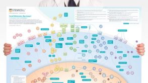 挂图Small Molecules, Big Impact in Pluripotent Stem Cell Research Overview of signaling pathways and small molecules in pluripotent stem cell research
 沪公网安备31010102008431号