Huber BC et al. (NOV 2013)
STEM CELLS 31 11 2354--2363
Costimulation-adhesion blockade is superior to Cyclosporine A and prednisone immunosuppressive therapy for preventing rejection of differentiated human embryonic stem cells following transplantation
RATIONALE: Human embryonic stem cell (hESC) derivatives are attractive candidates for therapeutic use. The engraftment and survival of hESC derivatives as xenografts or allografts require effective immunosuppression to prevent immune cell infiltration and graft destruction.backslashnbackslashnOBJECTIVE: To test the hypothesis that a short-course,dual-agent regimen of two costimulation-adhesion blockade agents can induce better engraftment of hESC derivatives compared to current immunosuppressive agents.backslashnbackslashnMETHODS AND RESULTS: We transduced hESCs with a double fusion reporter gene construct expressing firefly luciferase (Fluc) and enhanced green fluorescent protein,and differentiated these cells to endothelial cells (hESC-ECs). Reporter gene expression enabled longitudinal assessment of cell engraftment by bioluminescence imaging. Costimulation-adhesion therapy resulted in superior hESC-EC and mouse EC engraftment compared to cyclosporine therapy in a hind limb model. Costimulation-adhesion therapy also promoted robust hESC-EC and hESC-derived cardiomyocyte survival in an ischemic myocardial injury model. Improved hESC-EC engraftment had a cardioprotective effect after myocardial injury,as assessed by magnetic resonance imaging. Mechanistically,costimulation-adhesion therapy is associated with systemic and intragraft upregulation of T-cell immunoglobulin and mucin domain 3 (TIM3) and a reduced proinflammatory cytokine profile.backslashnbackslashnCONCLUSIONS: Costimulation-adhesion therapy is a superior alternative to current clinical immunosuppressive strategies for preventing the post-transplant rejection of hESC derivatives. By extending the window for cellular engraftment,costimulation-adhesion therapy enhances functional preservation following ischemic injury. This regimen may function through a TIM3-dependent mechanism.
View Publication
产品号#:
05850
05857
05870
05875
85850
85857
85870
85875
产品名:
mTeSR™1
mTeSR™1
Ban K et al. (OCT 2013)
Circulation 128 17 1897--1909
Purification of cardiomyocytes from differentiating pluripotent stem cells using molecular beacons that target cardiomyocyte-specific mRNA
BACKGROUND: Although methods for generating cardiomyocytes from pluripotent stem cells have been reported,current methods produce heterogeneous mixtures of cardiomyocytes and noncardiomyocyte cells. Here,we report an entirely novel system in which pluripotent stem cell-derived cardiomyocytes are purified by cardiomyocyte-specific molecular beacons (MBs). MBs are nanoscale probes that emit a fluorescence signal when hybridized to target mRNAs.backslashnbackslashnMETHOD AND RESULTS: Five MBs targeting mRNAs of either cardiac troponin T or myosin heavy chain 6/7 were generated. Among 5 MBs,an MB that targeted myosin heavy chain 6/7 mRNA (MHC1-MB) identified up to 99% of HL-1 cardiomyocytes,a mouse cardiomyocyte cell line,but textless3% of 4 noncardiomyocyte cell types in flow cytometry analysis,which indicates that MHC1-MB is specific for identifying cardiomyocytes. We delivered MHC1-MB into cardiomyogenically differentiated pluripotent stem cells through nucleofection. The detection rate of cardiomyocytes was similar to the percentages of cardiac troponin T- or cardiac troponin I-positive cardiomyocytes,which supports the specificity of MBs. Finally,MHC1-MB-positive cells were sorted by fluorescence-activated cell sorter from mouse and human pluripotent stem cell differentiating cultures,and ≈97% cells expressed cardiac troponin T or cardiac troponin I as determined by flow cytometry. These MB-based sorted cells maintained their cardiomyocyte characteristics,which was verified by spontaneous beating,electrophysiological studies,and expression of cardiac proteins. When transplanted in a myocardial infarction model,MB-based purified cardiomyocytes improved cardiac function and demonstrated significant engraftment for 4 weeks without forming tumors.backslashnbackslashnCONCLUSIONS: We developed a novel cardiomyocyte selection system that allows production of highly purified cardiomyocytes. These purified cardiomyocytes and this system can be valuable for cell therapy and drug discovery.
View Publication
产品号#:
05850
05857
05870
05875
85850
85857
85870
85875
产品名:
mTeSR™1
mTeSR™1
Qiu C et al. (FEB 2008)
Blood 111 4 2400--8
Globin switches in yolk sac-like primitive and fetal-like definitive red blood cells produced from human embryonic stem cells.
We have previously shown that coculture of human embryonic stem cells (hESCs) for 14 days with immortalized fetal hepatocytes yields CD34(+) cells that can be expanded in serum-free liquid culture into large numbers of megaloblastic nucleated erythroblasts resembling yolk sac-derived cells. We show here that these primitive erythroblasts undergo a switch in hemoglobin (Hb) composition during late terminal erythroid maturation with the basophilic erythroblasts expressing predominantly Hb Gower I (zeta(2)epsilon(2)) and the orthochromatic erythroblasts hemoglobin Gower II (alpha(2)epsilon(2)). This suggests that the switch from Hb Gower I to Hb Gower II,the first hemoglobin switch in humans is a maturation switch not a lineage switch. We also show that extending the coculture of the hESCs with immortalized fetal hepatocytes to 35 days yields CD34(+) cells that differentiate into more developmentally mature,fetal liver-like erythroblasts,that are smaller,express mostly fetal hemoglobin,and can enucleate. We conclude that hESC-derived erythropoiesis closely mimics early human development because the first 2 human hemoglobin switches are recapitulated,and because yolk sac-like and fetal liver-like cells are sequentially produced. Development of a method that yields erythroid cells with an adult phenotype remains necessary,because the most mature cells that can be produced with current systems express less than 2% adult beta-globin mRNA.
View Publication
产品号#:
09600
09650
18056
18056RF
产品名:
StemSpan™ SFEM
StemSpan™ SFEM
Porayette P et al. (DEC 2007)
Biochemical and biophysical research communications 364 3 522--527
Amyloid-?? precursor protein expression and modulation in human embryonic stem cells: A novel role for human chorionic gonadotropin
The amyloid-beta precursor protein (AbetaPP) is a ubiquitously expressed adhesion and neuritogenic protein whose processing has previously been shown to be regulated by reproductive hormones including the gonadotropin luteinizing hormone (LH) in human neuroblastoma cells. We report for the first time the expression of AbetaPP in human embryonic stem (hES) cells at the mRNA and protein levels. Using N- and C-terminal antibodies against AbetaPP,we detected both the mature and immature forms of AbetaPP as well as truncated variants ( approximately 53kDa,47kDa,and 29kDa) by immunoblot analyses. Expression of AbetaPP is regulated by both the stemness of the cells and pregnancy-associated hormones. Addition of human chorionic gonadotropin,the fetal equivalent of LH that is dramatically elevated during pregnancy,markedly increased the expression of all AbetaPP forms. These results indicate a critical molecular signaling link between the hormonal environment of pregnancy and the expression of AbetaPP in hES cells that is suggestive of an important function for this protein during early human embryogenesis prior to the formation of neural precursor cells.
View Publication
产品号#:
05850
05857
05870
05875
85850
85857
85870
85875
产品名:
mTeSR™1
mTeSR™1
Akutsu H et al. (JAN 2006)
Methods in enzymology 418 78--92
Human embryonic stem cells.
Human embryonic stem cells hold great promise in furthering our treatment of disease and increasing our understanding of early development. This chapter describes protocols for the derivation and maintenance of human embryonic stem cells. In addition,it summarizes briefly several alternative methods for the culture of human embryonic stem cells. Thus,this chapter provides a good starting point for researchers interested in harnessing the potential of human embryonic stem cells.
View Publication
产品号#:
05850
05857
05870
05875
85850
85857
85870
85875
产品名:
mTeSR™1
mTeSR™1
Kriz V et al. (NOV 2006)
The Journal of biological chemistry 281 45 34484--91
The SHB adapter protein is required for normal maturation of mesoderm during in vitro differentiation of embryonic stem cells.
Definitive mesoderm arises from a bipotent mesendodermal population,and to study processes controlling its development at this stage,embryonic stem (ES) cells can be employed. SHB (Src homology 2 protein in beta-cells) is an adapter protein previously found to be involved in ES cell differentiation to mesoderm. To further study the role of SHB in this context,we have established ES cell lines deficient for one (SHB+/-) or both SHB alleles (SHB-/-). Differentiating embryoid bodies (EBs) derived from these ES cell lines were used for gene expression analysis. Alternatively,EBs were stained for the blood vessel marker CD31. For hematopoietic differentiation,EBs were differentiated in methylcellulose. SHB-/- EBs exhibited delayed down-regulation of the early mesodermal marker Brachyury. Later mesodermal markers relatively specific for the hematopoietic,vascular,and cardiac lineages were expressed at lower levels on day 6 or 8 of differentiation in EBs lacking SHB. The expression of vascular endothelial growth factor receptor-2 and fibroblast growth factor receptor-1 was also reduced in SHB-/- EBs. SHB-/- EBs demonstrated impaired blood vessel formation after vascular endothelial growth factor stimulation. In addition,the SHB-/- ES cells formed fewer blood cell colonies than SHB+/+ ES cells. It is concluded that SHB is required for appropriate hematopoietic and vascular differentiation and that delayed down-regulation of Brachyury expression may play a role in this context.
View Publication
Yamashita J et al. (NOV 2000)
Nature 408 6808 92--6
Flk1-positive cells derived from embryonic stem cells serve as vascular progenitors.
Interaction between endothelial cells and mural cells (pericytes and vascular smooth muscle) is essential for vascular development and maintenance. Endothelial cells arise from Flk1-expressing (Flk1+) mesoderm cells,whereas mural cells are believed to derive from mesoderm,neural crest or epicardial cells and migrate to form the vessel wall. Difficulty in preparing pure populations of these lineages has hampered dissection of the mechanisms underlying vascular formation. Here we show that Flk1+ cells derived from embryonic stem cells can differentiate into both endothelial and mural cells and can reproduce the vascular organization process. Vascular endothelial growth factor promotes endothelial cell differentiation,whereas mural cells are induced by platelet-derived growth factor-BB. Vascular cells derived from Flk1+ cells can organize into vessel-like structures consisting of endothelial tubes supported by mural cells in three-dimensional culture. Injection of Flk1+ cells into chick embryos showed that they can incorporate as endothelial and mural cells and contribute to the developing vasculature in vivo. Our findings indicate that Flk1+ cells can act as 'vascular progenitor cells' to form mature vessels and thus offer potential for tissue engineering of the vascular system.
View Publication
产品号#:
06902
06952
00321
00322
00323
00324
00325
产品名:
C. L. Moreno et al. ( 2018)
Molecular neurodegeneration 13 1 33
BACKGROUND Type 2 diabetes (T2D) is a recognized risk factor for the development of cognitive impairment (CI) and/or dementia,although the exact nature of the molecular pathology of T2D-associated CI remains obscure. One link between T2D and CI might involve decreased insulin signaling in brain and/or neurons in either animal or postmortem human brains as has been reported as a feature of Alzheimer's disease (AD). Here we asked if neuronal insulin resistance is a cell autonomous phenomenon in a familial form of AD. METHODS We have applied a newly developed protocol for deriving human basal forebrain cholinergic neurons (BFCN) from skin fibroblasts via induced pluripotent stem cell (iPSC) technology. We generated wildtype and familial AD mutant PSEN2 N141I (presenilin 2) BFCNs and assessed if insulin signaling,insulin regulation of the major AD proteins Abeta$ and/or tau,and/or calcium fluxes is altered by the PSEN2 N141I mutation. RESULTS We report herein that wildtype,PSEN2 N141I and CRISPR/Cas9-corrected iPSC-derived BFCNs (and their precursors) show indistinguishable insulin signaling profiles as determined by the phosphorylation of canonical insulin signaling pathway molecules. Chronic insulin treatment of BFCNs of all genotypes led to a reduction in the Abeta$42/40 ratio. Unexpectedly,we found a CRISPR/Cas9-correctable effect of PSEN2 N141I on calcium flux,which could be prevented by chronic exposure of BFCNs to insulin. CONCLUSIONS Our studies indicate that the familial AD mutation PSEN2 N141I does not induce neuronal insulin resistance in a cell autonomous fashion. The ability of insulin to correct calcium fluxes and to lower Abeta$42/40 ratio suggests that insulin acts to oppose an AD-pathophysiology. Hence,our results are consistent with a potential physiological role for insulin as a mediator of resilience by counteracting specific metabolic and molecular features of AD.
View Publication

 EasySep™小鼠TIL(CD45)正选试剂盒


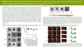 科学海报STEMdiff™ Cerebral Organoid Kit: A New Tool for the Culture of 3D Brain Organoids Derived from hPSCs

 沪公网安备31010102008431号