Freude KK et al. (JUL 2011)
Journal of Biological Chemistry 286 27 24264--24274
Soluble amyloid precursor protein induces rapid neural differentiation of human embryonic stem cells.
Human embryonic stem cells (hESCs) offer tremendous potential for not only treating neurological disorders but also for their ability to serve as vital reagents to model and investigate human disease. To further our understanding of a key protein involved in Alzheimer disease pathogenesis,we stably overexpressed amyloid precursor protein (APP) in hESCs. Remarkably,we found that APP overexpression in hESCs caused a rapid and robust differentiation of pluripotent stem cells toward a neural fate. Despite maintenance in standard hESC media,up to 80% of cells expressed the neural stem cell marker nestin,and 65% exhibited the more mature neural marker β-3 tubulin within just 5 days of passaging. To elucidate the mechanism underlying the effects of APP on neural differentiation,we examined the proteolysis of APP and performed both gain of function and loss of function experiments. Taken together,our results demonstrate that the N-terminal secreted soluble forms of APP (in particular sAPPβ) robustly drive neural differentiation of hESCs. Our findings not only reveal a novel and intriguing role for APP in neural lineage commitment but also identify a straightforward and rapid approach to generate large numbers of neurons from human embryonic stem cells. These novel APP-hESC lines represent a valuable tool to investigate the potential role of APP in development and neurodegeneration and allow for insights into physiological functions of this protein.
View Publication
产品号#:
05850
05857
05870
05875
85850
85857
85870
85875
产品名:
mTeSR™1
mTeSR™1
Batista LFZ et al. (JUN 2011)
Nature 474 7351 399--402
Telomere shortening and loss of self-renewal in dyskeratosis congenita induced pluripotent stem cells
The differentiation of patient-derived induced pluripotent stem cells (iPSCs) to committed fates such as neurons,muscle and liver is a powerful approach for understanding key parameters of human development and disease. Whether undifferentiated iPSCs themselves can be used to probe disease mechanisms is uncertain. Dyskeratosis congenita is characterized by defective maintenance of blood,pulmonary tissue and epidermal tissues and is caused by mutations in genes controlling telomere homeostasis. Short telomeres,a hallmark of dyskeratosis congenita,impair tissue stem cell function in mouse models,indicating that a tissue stem cell defect may underlie the pathophysiology of dyskeratosis congenita. Here we show that even in the undifferentiated state,iPSCs from dyskeratosis congenita patients harbour the precise biochemical defects characteristic of each form of the disease and that the magnitude of the telomere maintenance defect in iPSCs correlates with clinical severity. In iPSCs from patients with heterozygous mutations in TERT,the telomerase reverse transcriptase,a 50% reduction in telomerase levels blunts the natural telomere elongation that accompanies reprogramming. In contrast,mutation of dyskerin (DKC1) in X-linked dyskeratosis congenita severely impairs telomerase activity by blocking telomerase assembly and disrupts telomere elongation during reprogramming. In iPSCs from a form of dyskeratosis congenita caused by mutations in TCAB1 (also known as WRAP53),telomerase catalytic activity is unperturbed,yet the ability of telomerase to lengthen telomeres is abrogated,because telomerase mislocalizes from Cajal bodies to nucleoli within the iPSCs. Extended culture of DKC1-mutant iPSCs leads to progressive telomere shortening and eventual loss of self-renewal,indicating that a similar process occurs in tissue stem cells in dyskeratosis congenita patients. These findings in iPSCs from dyskeratosis congenita patients reveal that undifferentiated iPSCs accurately recapitulate features of a human stem cell disease and may serve as a cell-culture-based system for the development of targeted therapeutics.
View Publication
产品号#:
05850
05857
05870
05875
85850
85857
85870
85875
产品名:
mTeSR™1
mTeSR™1
Kallas A et al. (APR 2011)
PLoS ONE 6 4 e19114
Nocodazole treatment decreases expression of pluripotency markers nanog and Oct4 in human embryonic stem cells
Nocodazole is a known destabiliser of microtubule dynamics and arrests cell-cycle at the G2/M phase. In the context of the human embryonic stem cell (hESC) it is important to understand how this arrest influences the pluripotency of cells. Here we report for the first time the changes in the expression of transcription markers Nanog and Oct4 as well as SSEA-3 and SSEA-4 in human embryonic cells after their treatment with nocodazole. Multivariate permeabilised-cell flow cytometry was applied for characterising the expression of Nanog and Oct4 during different cell cycle phases. Among untreated hESC we detected Nanog-expressing cells,which also expressed Oct4,SSEA-3 and SSEA-4. We also found another population expressing SSEA-4,but without Nanog,Oct4 and SSEA-3 expression. Nocodazole treatment resulted in a decrease of cell population positive for all four markers Nanog,Oct4,SSEA-3,SSEA-4. Nocodazole-mediated cell-cycle arrest was accompanied by higher rate of apoptosis and upregulation of p53. Twenty-four hours after the release from nocodazole block,the cell cycle of hESC normalised,but no increase in the expression of transcription markers Nanog and Oct4 was detected. In addition,the presence of ROCK-2 inhibitor Y-27632 in the medium had no effect on increasing the expression of pluripotency markers Nanog and Oct4 or decreasing apoptosis or the level of p53. The expression of SSEA-3 and SSEA-4 increased in Nanog-positive cells after wash-out of nocodazole in the presence and in the absence of Y-27632. Our data show that in hESC nocodazole reversible blocks cell cycle,which is accompanied by irreversible loss of expression of pluripotency markers Nanog and Oct4.
View Publication
产品号#:
05850
05857
05870
05875
85850
85857
85870
85875
产品名:
mTeSR™1
mTeSR™1
Chen G et al. (MAY 2011)
Nature methods 8 5 424--9
Chemically defined conditions for human iPSC derivation and culture.
We re-examine the individual components for human embryonic stem cell (ESC) and induced pluripotent stem cell (iPSC) culture and formulate a cell culture system in which all protein reagents for liquid media,attachment surfaces and splitting are chemically defined. A major improvement is the lack of a serum albumin component,as variations in either animal- or human-sourced albumin batches have previously plagued human ESC and iPSC culture with inconsistencies. Using this new medium (E8) and vitronectin-coated surfaces,we demonstrate improved derivation efficiencies of vector-free human iPSCs with an episomal approach. This simplified E8 medium should facilitate both the research use and clinical applications of human ESCs and iPSCs and their derivatives,and should be applicable to other reprogramming methods.
View Publication
Pluripotent male germline stem cells from goat fetal testis and their survival in mouse testis.
Male germline stem cells (mGSCs) are stem cells present in male testis responsible for spermatogenesis during their whole life. Studies have shown that mGSCs can be derived in vitro and resemble embryonic stem cells (ESCs) properties both in the mouse and humans. However,little is know about these cells in domestic animals. Here we report the first successful establishment of goat GSCs derived from 2-5-month fetal testis,and developmental potential assay of these cells both in vitro and in vivo. These cells express pluripotent markers such as Oct4,Sox2,C-myc,and Tert when cultured as human ESCs conditions. Embryoid bodies (EBs) formed by goat mGSCs were induced with 2 × 10(-6) M retinoic acid (RA). Immunofluorescence analysis showed that some cells inside of the EBs were positive for meiosis marker-SCP3,STRA8,and germ cell marker-VASA,and haploid marker-FE-J1,PRM1,indicating their germ cell lineage differentiation. Some cells become elongated sperm-like cells after induction. Approximately 34.88% (30/86) embryos showed cleavage and four embryos were cultured on murine fibroblast feeder and formed small embryonic stem like colonies. However,most stalled at four-cell stage after intracytoplasmic sperm injection (ICSI) of these cells. Transplantation of DAPI labeled mGSCs into the seminiferous tubules of busulfan-treated mice,and showed that mGSCs can colonize,self-renew,and differentiate into germ cells. Thus,we have established a goat GSC cell line and these cells could be differentiated into sperm-like cells in vivo and sperms in vitro,providing a promising platform for generation of transgenic goat for production of specific humanized proteins.
View Publication
产品号#:
05850
05857
05870
05875
85850
85857
85870
85875
产品名:
mTeSR™1
mTeSR™1
Bak XY et al. (NOV 2011)
Human gene therapy 22 11 1365--77
Human embryonic stem cell-derived mesenchymal stem cells as cellular delivery vehicles for prodrug gene therapy of glioblastoma.
Mesenchymal stem cells (MSCs) possess tumor-tropic properties and consequently have been used to deliver therapeutic agents for cancer treatment. Their potential in cancer therapy highlights the need for a consistent and renewable source for the production of uniform human MSCs suitable for clinical applications. In this study,we seek to investigate whether human embryonic stem cells can be used as a cell source to fulfill this goal. We generated MSC-like cells from two human embryonic stem cell lines,HuES9 and H1,and observed that MSC-like cells derived from human embryonic stem cells were able to migrate into human glioma intracranial xenografts after being injected into the cerebral hemisphere contralateral to the tumor inoculation site. We engineered these cells with baculoviral and lentiviral vectors,respectively,for transient and stable expression of the herpes simplex virus thymidine kinase gene. In tumor-bearing mice the engineered MSC-like cells were capable of inhibiting tumor growth and prolonging survival in the presence of ganciclovir after they were injected either directly into the xenografts or into the opposite hemisphere. Our findings suggest that human embryonic stem cell-derived MSCs may be a viable and attractive alternative for large-scale derivation of targeting vehicles for cancer therapy.
View Publication
Nishimura K et al. (FEB 2011)
The Journal of biological chemistry 286 6 4760--71
Development of defective and persistent Sendai virus vector: a unique gene delivery/expression system ideal for cell reprogramming.
The ectopic expression of transcription factors can reprogram differentiated tissue cells into induced pluripotent stem cells. However,this is a slow and inefficient process,depending on the simultaneous delivery of multiple genes encoding essential reprogramming factors and on their sustained expression in target cells. Moreover,once cell reprogramming is accomplished,these exogenous reprogramming factors should be replaced with their endogenous counterparts for establishing autoregulated pluripotency. Complete and designed removal of the exogenous genes from the reprogrammed cells would be an ideal option for satisfying this latter requisite as well as for minimizing the risk of malignant cell transformation. However,no single gene delivery/expression system has ever been equipped with these contradictory characteristics. Here we report the development of a novel replication-defective and persistent Sendai virus (SeVdp) vector based on a noncytopathic variant virus,which fulfills all of these requirements for cell reprogramming. The SeVdp vector could accommodate up to four exogenous genes,deliver them efficiently into various mammalian cells (including primary tissue cells and human hematopoietic stem cells) and express them stably in the cytoplasm at a prefixed balance. Furthermore,interfering with viral transcription/replication using siRNA could erase the genomic RNA of SeVdp vector from the target cells quickly and thoroughly. A SeVdp vector installed with Oct4/Sox2/Klf4/c-Myc could reprogram mouse primary fibroblasts quite efficiently; ∼1% of the cells were reprogrammed to Nanog-positive induced pluripotent stem cells without chromosomal gene integration. Thus,this SeVdp vector has potential as a tool for advanced cell reprogramming and for stem cell research.
View Publication
产品号#:
产品名:
Stumpf M et al. (DEC 2010)
Proceedings of the National Academy of Sciences of the United States of America 107 50 21541--6
Specific erythroid-lineage defect in mice conditionally deficient for Mediator subunit Med1.
The Mediator complex forms the bridge between transcriptional activators and the RNA polymerase II. Med1 (also known as PBP or TRAP220) is a key component of Mediator that interacts with nuclear hormone receptors and GATA transcription factors. Here,we show dynamic recruitment of GATA-1,TFIIB,Mediator,and RNA polymerase II to the β-globin locus in induced mouse erythroid leukemia cells and in an erythropoietin-inducible hematopoietic progenitor cell line. Using Med1 conditional knockout mice,we demonstrate a specific block in erythroid development but not in myeloid or lymphoid development,highlighted by the complete absence of β-globin gene expression. Thus,Mediator subunit Med1 plays a pivotal role in erythroid development and in β-globin gene activation.
View Publication
Nekrasov ED et al. (DEC 2016)
Molecular Neurodegeneration 11 1 1--15
Manifestation of Huntington's disease pathology in human induced pluripotent stem cell-derived neurons.
Background: Huntington's disease (HD) is an incurable hereditary neurodegenerative disorder,which manifests itself as a loss of GABAergic medium spiny (GABA MS) neurons in the striatum and caused by an expansion of the CAG repeat in exon 1 of the huntingtin gene. There is no cure for HD,existing pharmaceutical can only relieve its symptoms. Results: Here,induced pluripotent stem cells were established from patients with low CAG repeat expansion in the huntingtin gene,and were then efficiently differentiated into GABA MS-like neurons (GMSLNs) under defined culture conditions. The generated HD GMSLNs recapitulated disease pathology in vitro,as evidenced by mutant huntingtin protein aggregation,increased number of lysosomes/autophagosomes,nuclear indentations,and enhanced neuronal death during cell aging. Moreover,store-operated channel (SOC) currents were detected in the differentiated neurons,and enhanced calcium entry was reproducibly demonstrated in all HD GMSLNs genotypes. Additionally,the quinazoline derivative,EVP4593,reduced the number of lysosomes/autophagosomes and SOC currents in HD GMSLNs and exerted neuroprotective effects during cell aging. Conclusions: Our data is the first to demonstrate the direct link of nuclear morphology and SOC calcium deregulation to mutant huntingtin protein expression in iPSCs-derived neurons with disease-mimetic hallmarks,providing a valuable tool for identification of candidate anti-HD drugs. Our experiments demonstrated that EVP4593 may be a promising anti-HD drug. [ABSTRACT FROM AUTHOR]
View Publication
产品号#:
05854
05855
05850
05857
05870
05875
85850
85857
85870
85875
产品名:
mFreSR™
mFreSR™
mTeSR™1
mTeSR™1
Christoffersson J et al. (APR 2016)
Methods in molecular biology (Clifton,N.J.)
A Microfluidic Bioreactor for Toxicity Testing of Stem Cell Derived 3D Cardiac Bodies.
Modeling tissues and organs using conventional 2D cell cultures is problematic as the cells rapidly lose their in vivo phenotype. In microfluidic bioreactors the cells reside in microstructures that are continuously perfused with cell culture medium to provide a dynamic environment mimicking the cells natural habitat. These micro scale bioreactors are sometimes referred to as organs-on-chips and are developed in order to improve and extend cell culture experiments. Here,we describe the two manufacturing techniques photolithography and soft lithography that are used in order to easily produce microfluidic bioreactors. The use of these bioreactors is exemplified by a toxicity assessment on 3D clustered human pluripotent stem cells (hPSC)-derived cardiomyocytes by beating frequency imaging.
View Publication

 EasySep™小鼠TIL(CD45)正选试剂盒


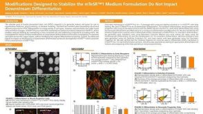 科学海报Modifications Designed to Stabilize the mTeSR1™ Formulation Do Not Impact Downstream Differentiation
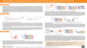 科学海报A Reproducible and Simple Method to Generate Red Blood Cells From Human Pluripotent Stem Cells
 沪公网安备31010102008431号