Kong C-W et al. (MAR 2017)
Stem cell research 19 76--81
Increasing the physical size and nucleation status of human pluripotent stem cell-derived ventricular cardiomyocytes by cell fusion.
Human pluripotent stem cell-derived cardiomyocytes (hPSC-CMs) provide an unlimited source of donor cells for potential cardiac regenerative therapies. However,hPSC-CMs are immature. For instance,hPSC-CMs are only 1/10 of the physical size of their adult counterparts; the majority are mono- rather than bi- or multi-nucleated,which is an evolutionary adaptive feature in metabolically active cells such as adult CMs. Here,we attempted to increase the physical size and nucleation status of hPSC-derived ventricular (V) cardiomyocytes (hPSC-VCMs) using chemically-induced cell fusion,and examined the subsequent functional effects. Polyethylene glycol (PEG) was employed to fuse a 1:1 mixture of lentiviral vectors LV-MLC2v-GFP- or -tdTomato-labeled hPSC-VCMs,such that hPSC-VCMs fused syncytia (FS) were identified as doubly GFP(+)/tdTomato(+) multi-nucleated cells. These microscopically-identified FS were doubled in size as gauged by their capacitance when compared to the control mononucleated hPSC-VCMs using patch-clamp analysis. Reduced automaticity or action potential (AP) firing rate and moderately prolonged AP duration were observed in FS from day 6 post-fusion induction. However,Ca(2+) handling,mitochondrial biogenesis and the extent of apoptosis were not significantly altered. We conclude that larger,multi-nucleated hPSC-VCMs FS can be created by chemically-induced cell fusion but global maturation requires additional triggering cues.
View Publication
Chemically defined generation of human cardiomyocytes.
Existing methods for human induced pluripotent stem cell (hiPSC) cardiac differentiation are efficient but require complex,undefined medium constituents that hinder further elucidation of the molecular mechanisms of cardiomyogenesis. Using hiPSCs derived under chemically defined conditions on synthetic matrices,we systematically developed an optimized cardiac differentiation strategy,using a chemically defined medium consisting of just three components: the basal medium RPMI 1640,L-ascorbic acid 2-phosphate and rice-derived recombinant human albumin. Along with small molecule-based induction of differentiation,this protocol produced contractile sheets of up to 95% TNNT2(+) cardiomyocytes at a yield of up to 100 cardiomyocytes for every input pluripotent cell and was effective in 11 hiPSC lines tested. This chemically defined platform for cardiac specification of hiPSCs will allow the elucidation of cardiomyocyte macromolecular and metabolic requirements and will provide a minimal system for the study of maturation and subtype specification.
View Publication
产品号#:
05850
05857
05870
05875
85850
85857
85870
85875
产品名:
mTeSR™1
mTeSR™1
Dambrot C et al. (AUG 2014)
Journal of Cellular and Molecular Medicine 18 8 1509--1518
Serum supplemented culture medium masks hypertrophic phenotypes in human pluripotent stem cell derived cardiomyocytes
It has been known for over 20 years that foetal calf serum can induce hypertrophy in cultured cardiomyocytes but this is rarely considered when examining cardiomyocytes derived from pluripotent stem cells (PSC). Here,we determined how serum affected cardiomyocytes from human embryonic- (hESC) and induced pluripotent stem cells (hiPSC) and hiPSC from patients with hypertrophic cardiomyopathy linked to a mutation in the MYBPC3 gene. We first confirmed previously published hypertrophic effects of serum on cultured neonatal rat cardiomyocytes demonstrated as increased cell surface area and beating frequency. We then found that serum increased the cell surface area of hESC- and hiPSC-derived cardiomyocytes and their spontaneous contraction rate. Phenylephrine,which normally induces cardiac hypertrophy,had no additional effects under serum conditions. Likewise,hiPSC-derived cardiomyocytes from three MYBPC3 patients which had a greater surface area than controls in the absence of serum as predicted by their genotype,did not show this difference in the presence of serum. Serum can thus alter the phenotype of human PSC derived cardiomyocytes under otherwise defined conditions such that the effects of hypertrophic drugs and gene mutations are underestimated. It is therefore pertinent to examine cardiac phenotypes in culture media without or in low concentrations of serum.
View Publication
产品号#:
05850
05857
05870
05875
85850
85857
85870
85875
产品名:
mTeSR™1
mTeSR™1
Castro-Diaz N et al. (JUL 2014)
Genes and Development 28 13 1397--1409
Evolutionally dynamic L1 regulation in embryonic stem cells
Mobile elements are important evolutionary forces that challenge genomic integrity. Long interspersed element-1 (L1,also known as LINE-1) is the only autonomous transposon still active in the human genome. It displays an unusual pattern of evolution,with,at any given time,a single active L1 lineage amplifying to thousands of copies before getting replaced by a new lineage,likely under pressure of host restriction factors,which act notably by silencing L1 expression during early embryogenesis. Here,we demonstrate that in human embryonic stem (hES) cells,KAP1 (KRAB [Kruppel-associated box domain]-associated protein 1),the master cofactor of KRAB-containing zinc finger proteins (KRAB-ZFPs) previously implicated in the restriction of endogenous retroviruses,represses a discrete subset of L1 lineages predicted to have entered the ancestral genome between 26.8 million and 7.6 million years ago. In mice,we documented a similar chronologically conditioned pattern,albeit with a much contracted time scale. We could further identify an L1-binding KRAB-ZFP,suggesting that this rapidly evolving protein family is more globally responsible for L1 recognition. KAP1 knockdown in hES cells induced the expression of KAP1-bound L1 elements,but their younger,human-specific counterparts (L1Hs) were unaffected. Instead,they were stimulated by depleting DNA methyltransferases,consistent with recent evidence demonstrating that the PIWI-piRNA (PIWI-interacting RNA) pathway regulates L1Hs in hES cells. Altogether,these data indicate that the early embryonic control of L1 is an evolutionarily dynamic process and support a model in which newly emerged lineages are first suppressed by DNA methylation-inducing small RNA-based mechanisms before KAP1-recruiting protein repressors are selected.
View Publication
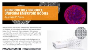
A 3D sphere culture system containing functional polymers for large-scale human pluripotent stem cell production
Utilizing human pluripotent stem cells (hPSCs) in cell-based therapy and drug discovery requires large-scale cell production. However,scaling up conventional adherent cultures presents challenges of maintaining a uniform high quality at low cost. In this regard,suspension cultures are a viable alternative,because they are scalable and do not require adhesion surfaces. 3D culture systems such as bioreactors can be exploited for large-scale production. However,the limitations of current suspension culture methods include spontaneous fusion between cell aggregates and suboptimal passaging methods by dissociation and reaggregation. 3D culture systems that dynamically stir carrier beads or cell aggregates should be refined to reduce shearing forces that damage hPSCs. Here,we report a simple 3D sphere culture system that incorporates mechanical passaging and functional polymers. This setup resolves major problems associated with suspension culture methods and dynamic stirring systems and may be optimal for applications involving large-scale hPSC production. ?? 2014 The Authors.
View Publication
产品号#:
05850
05857
05870
05875
85850
85857
85870
85875
产品名:
mTeSR™1
mTeSR™1
Mormone E et al. (NOV 2014)
Stem cells and development 23 21 2626--36
Footprint-free" human induced pluripotent stem cell-derived astrocytes for in vivo cell-based therapy."
The generation of human induced pluripotent stem cells (hiPSC) from somatic cells has enabled the possibility to provide patient-specific hiPSC for cell-based therapy,drug discovery,and other translational applications. Two major obstacles in using hiPSC for clinical application reside in the risk of genomic modification when they are derived with viral transgenes and risk of teratoma formation if undifferentiated cells are engrafted. In this study,we report the generation of footprint-free" hiPSC-derived astrocytes. These are efficiently generated�
View Publication
产品号#:
05850
05857
05870
05875
85850
85857
85870
85875
产品名:
mTeSR™1
mTeSR™1
Liu Y et al. (MAR 2015)
Journal of Biomedical Materials Research - Part A 103 3 1053--1059
Native nucleus pulposus tissue matrix promotes notochordal differentiation of human induced pluripotent stem cells with potential for treating intervertebral disc degeneration
Native porcine nucleus pulposus (NP) tissue harbors a number of notochordal cells (NCs). Whether the native NP matrix supports the homeostasis of notochordal cells is poorly understood. We hypothesized the NP matrix alone may contain sufficient regulatory factors and can serve as stimuli to generate notochordal cells (NCs) from human pluripotent stem cells. NCs are a promising cell sources for cell-based therapy to treat some types of intervertebral disc (IVD) degeneration. One major limitation of this emerging technique is the lack of available NCs as a potential therapeutic cell source. Human pluripotent stem cells derived from reprogramming or somatic cell nuclear transfer technique may yield stable and unlimited source for therapeutic use. We devised a new method to use porcine NP matrix to direct notochordal differentiation of human induced pluripotent stem cells (hiPSCs). The results showed that hiPSCs successfully differentiated into NC-like cells under the influence of devitalized porcine NP matrix. The NC-like cells expressed typical notochordal marker genes including brachyury (T),cytokeratin-8 (CK-8) and cytokeratin-18 (CK-18),and they displayed the ability to generate NP-like tissue in vitro,which was rich in aggrecan and collagen type II. These findings demonstrated the proof of concept for using native NP matrix to direct notochordal differentiation of hiPSCs. It provides a foundation for further understanding the biology of NCs,and eventually towards regenerative therapies for disc degeneration.
View Publication
产品号#:
05850
05857
05870
05875
85850
85857
85870
85875
产品名:
mTeSR™1
mTeSR™1
Baatz JE et al. (JUL 2014)
In vivo (Athens,Greece) 28 4 411--423
Cryopreservation of viable human lung tissue for versatile post-thaw analyses and culture.
Clinical trials are currently used to test therapeutic efficacies for lung cancer,infections and diseases. Animal models are also used as surrogates for human disease. Both approaches are expensive and time-consuming. The utility of human biospecimens as models is limited by specialized tissue processing methods that preserve subclasses of analytes (e.g. RNA,protein,morphology) at the expense of others. We present a rapid and reproducible method for the cryopreservation of viable lung tissue from patients undergoing lobectomy or transplant. This method involves the pseudo-diaphragmatic expansion of pieces of fresh lung tissue with cryoprotectant formulation (pseudo-diaphragmatic expansion-cryoprotectant perfusion or PDX-CP) followed by controlled-rate freezing in cryovials. Expansion-perfusion rates,volumes and cryoprotectant formulation were optimized to maintain tissue architecture,decrease crystal formation and increase long-term cell viability. Rates of expansion of 4 cc/min or less and volumes ranging from 0.8-1.2 × tissue volume were well-tolerated by lung tissue obtained from patients with chronic obstructive pulmonary disease or idiopathic pulmonary fibrosis,showing minimal differences compared to standard histopathology. Morphology was greatly improved by the PDX-CP procedure compared to simple fixation. Fresh versus post-thawed lung tissue showed minimal differences in histology,RNA integrity numbers and post-translational modified protein integrity (2-dimensional differential gel electrophoresis). It was possible to derive numerous cell types,including alveolar epithelial cells,fibroblasts and stem cells,from the tissue for at least three months after cryopreservation. This new method should provide a uniform,cost-effective approach to the banking of biospecimens,with versatility to be amenable to any post-acquisition process applicable to fresh tissue samples.
View Publication

 EasySep™小鼠TIL(CD45)正选试剂盒


 挂图Small Molecules, Big Impact in Pluripotent Stem Cell Research Overview of signaling pathways and small molecules in pluripotent stem cell research
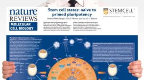 挂图Stem Cell States: Naive to Primed Pluripotency Properties of naive (ground) and primed pluripotent stem cells

 沪公网安备31010102008431号