Induced pluripotent stem cells with a mitochondrial dna deletion
In congenital mitochondrial DNA (mtDNA) disorders,a mixture of normal and mutated mtDNA (termed heteroplasmy) exists at varying levels in different tissues,which determines the severity and phenotypic expression of disease. Pearson marrow pancreas syndrome (PS) is a congenital bone marrow failure disorder caused by heteroplasmic deletions in mtDNA. The cause of the hematopoietic failure in PS is unknown,and adequate cellular and animal models are lacking. Induced pluripotent stem (iPS) cells are particularly amenable for studying mtDNA disorders,as cytoplasmic genetic material is retained during direct reprogramming. Here,we derive and characterize iPS cells from a patient with PS. Taking advantage of the tendency for heteroplasmy to change with cell passage,we isolated isogenic PS-iPS cells without detectable levels of deleted mtDNA. We found that PS-iPS cells carrying a high burden of deleted mtDNA displayed differences in growth,mitochondrial function,and hematopoietic phenotype when differentiated in vitro,compared to isogenic iPS cells without deleted mtDNA. Our results demonstrate that reprogramming somatic cells from patients with mtDNA disorders can yield pluripotent stem cells with varying burdens of heteroplasmy that might be useful in the study and treatment of mitochondrial diseases. STEM CELLS2013;31:1287–1297
View Publication
产品号#:
04434
04444
05850
05857
05870
05875
07923
85850
85857
85870
85875
产品名:
MethoCult™ H4434 Classic
MethoCult™ H4434 Classic
Dispase (1 U/mL)
mTeSR™1
mTeSR™1
Awe JP et al. (JUL 2013)
Stem cell research & therapy 4 4 87
Generation and characterization of transgene-free human induced pluripotent stem cells and conversion to putative clinical-grade status
INTRODUCTION: The reprogramming of a patient's somatic cells back into induced pluripotent stem cells (iPSCs) holds significant promise for future autologous cellular therapeutics. The continued presence of potentially oncogenic transgenic elements following reprogramming,however,represents a safety concern that should be addressed prior to clinical applications. The polycistronic stem cell cassette (STEMCCA),an excisable lentiviral reprogramming vector,provides,in our hands,the most consistent reprogramming approach that addresses this safety concern. Nevertheless,most viral integrations occur in genes,and exactly how the integration,epigenetic reprogramming,and excision of the STEMCCA reprogramming vector influences those genes and whether these cells still have clinical potential are not yet known. METHODS: In this study,we used both microarray and sensitive real-time PCR to investigate gene expression changes following both intron-based reprogramming and excision of the STEMCCA cassette during the generation of human iPSCs from adult human dermal fibroblasts. Integration site analysis was conducted using nonrestrictive linear amplification PCR. Transgene-free iPSCs were fully characterized via immunocytochemistry,karyotyping and teratoma formation,and current protocols were implemented for guided differentiation. We also utilized current good manufacturing practice guidelines and manufacturing facilities for conversion of our iPSCs into putative clinical grade conditions. RESULTS: We found that a STEMCCA-derived iPSC line that contains a single integration,found to be located in an intronic location in an actively transcribed gene,PRPF39,displays significantly increased expression when compared with post-excised stem cells. STEMCCA excision via Cre recombinase returned basal expression levels of PRPF39. These cells were also shown to have proper splicing patterns and PRPF39 gene sequences. We also fully characterized the post-excision iPSCs,differentiated them into multiple clinically relevant cell types (including oligodendrocytes,hepatocytes,and cardiomyocytes),and converted them to putative clinical-grade conditions using the same approach previously approved by the US Food and Drug Administration for the conversion of human embryonic stem cells from research-grade to clinical-grade status. CONCLUSION: For the first time,these studies provide a proof-of-principle for the generation of fully characterized transgene-free human iPSCs and,in light of the limited availability of current good manufacturing practice cellular manufacturing facilities,highlight an attractive potential mechanism for converting research-grade cell lines into putatively clinical-grade biologics for personalized cellular therapeutics.
View Publication
Kearns NA et al. (NOV 2013)
Stem Cell Research 11 3 1003--1012
Generation of organized anterior foregut epithelia from pluripotent stem cells using small molecules
Anterior foregut endoderm (AFE) gives rise to therapeutically relevant cell types in tissues such as the esophagus,salivary glands,lung,thymus,parathyroid and thyroid. Despite its importance,reports describing the generation of AFE from pluripotent stem cells (PSCs) by directed differentiation have mainly focused on the Nkx2.1(+) lung and thyroid lineages. Here,we describe a novel protocol to derive a subdomain of AFE,identified by expression of Pax9,from PSCs using small molecules and defined media conditions. We generated a reporter PSC line for isolation and characterization of Pax9(+) AFE cells,which when transplanted in vivo,can form several distinct complex AFE-derived epithelia,including mucosal glands and stratified squamous epithelium. Finally,we show that the directed differentiation protocol can be used to generate AFE from human PSCs. Thus,this work both broadens the range of PSC-derived AFE tissues and creates a platform enabling the study of AFE disorders.
View Publication
Malik J et al. (NOV 2013)
Haematologica 98 11 1778--1787
Erythropoietin critically regulates the terminal maturation of murine and human primitive erythroblasts
Primitive erythroid cells,the first red blood cells produced in the mammalian embryo,are necessary for embryonic survival. Erythropoietin and its receptor EpoR,are absolutely required for survival of late-stage definitive erythroid progenitors in the fetal liver and adult bone marrow. Epo- and Epor-null mice die at E13.5 with a lack of definitive erythrocytes. However,the persistence of circulating primitive erythroblasts raises questions about the role of erythropoietin/EpoR in primitive erythropoiesis. Using Epor-null mice and a novel primitive erythroid 2-step culture we found that erythropoietin is not necessary for specification of primitive erythroid progenitors. However,Epor-null embryos develop a progressive,profound anemia by E12.5 as primitive erythroblasts mature as a synchronous cohort. This anemia results from reduced primitive erythroblast proliferation associated with increased p27 expression,from advanced cellular maturation,and from markedly elevated rates of apoptosis associated with an imbalance in pro- and anti-apoptotic gene expression. Both mouse and human primitive erythroblasts cultured without erythropoietin also undergo accelerated maturation and apoptosis at later stages of maturation. We conclude that erythropoietin plays an evolutionarily conserved role in promoting the proliferation,survival,and appropriate timing of terminal maturation of primitive erythroid precursors.
View Publication
产品号#:
05850
05857
05870
05875
85850
85857
85870
85875
05270
05275
产品名:
mTeSR™1
mTeSR™1
STEMdiff™ APEL™2 培养基
STEMdiff™ APEL™2 培养基
Burkhardt MF et al. (SEP 2013)
Molecular and Cellular Neuroscience 56 355--364
A cellular model for sporadic ALS using patient-derived induced pluripotent stem cells
Development of therapeutics for genetically complex neurodegenerative diseases such as sporadic amyotrophic lateral sclerosis (ALS) has largely been hampered by lack of relevant disease models. Reprogramming of sporadic ALS patients' fibroblasts into induced pluripotent stem cells (iPSC) and differentiation into affected neurons that show a disease phenotype could provide a cellular model for disease mechanism studies and drug discovery. Here we report the reprogramming to pluripotency of fibroblasts from a large cohort of healthy controls and ALS patients and their differentiation into motor neurons. We demonstrate that motor neurons derived from three sALS patients show de novo TDP-43 aggregation and that the aggregates recapitulate pathology in postmortem tissue from one of the same patients from which the iPSC were derived. We configured a high-content chemical screen using the TDP-43 aggregate endpoint both in lower motor neurons and upper motor neuron like cells and identified FDA-approved small molecule modulators including Digoxin demonstrating the feasibility of patient-derived iPSC-based disease modeling for drug screening.
View Publication
产品号#:
05850
05857
05870
05875
85850
85857
85870
85875
产品名:
mTeSR™1
mTeSR™1
van der Meer AD et al. (SEP 2013)
Lab on a Chip 13 18 3562--3568
Three-dimensional co-cultures of human endothelial cells and embryonic stem cell-derived pericytes inside a microfluidic device
Organs-on-chips are microengineered in vitro tissue structures that can be used as platforms for physiological and pathological research. They provide tissue-like microenvironments in which different cell types can be co-cultured in a controlled manner to create synthetic organ mimics. Blood vessels are an integral part of all tissues in the human body. Development of vascular structures is therefore an important research topic for advancing the field of organs-on-chips since generated tissues will require a blood or nutrient supply. Here,we have engineered three-dimensional constructs of vascular tissue inside microchannels by injecting a mixture of human umbilical vein endothelial cells,human embryonic stem cell-derived pericytes (the precursors of vascular smooth muscle cells) and rat tail collagen I into a polydimethylsiloxane microfluidic channel with dimensions 500 μm × 120 μm × 1 cm (w × h × l). Over the course of 12 h,the cells organized themselves into a single long tube resembling a blood vessel that followed the contours of the channel. Detailed examination of tube morphology by confocal microscopy revealed a mature endothelial monolayer with complete PECAM-1 staining at cell–cell contacts and pericytes incorporated inside the tubular structures. We also demonstrated that tube formation was disrupted in the presence of a neutralizing antibody against transforming growth factor-beta (TGF-β). The TGF-β signaling pathway is essential for normal vascular development; deletion of any of its components in mouse development results in defective vasculogenesis and angiogenesis and mutations in humans have been linked to multiple vascular genetic diseases. In the engineered microvessels,inhibition of TGF-β signaling resulted in tubes with smaller diameters and higher tortuosity,highly reminiscent of the abnormal vessels observed in patients with one particular vascular disease known as hereditary hemorrhagic telangiectasia (HHT). In summary,we have developed microengineered three-dimensional vascular structures that can be used as a model to test the effects of drugs and study the interaction between different human vascular cell types. In the future,the model may be integrated into larger tissue constructs to advance the development of organs-on-chips.
View Publication

 EasySep™小鼠TIL(CD45)正选试剂盒


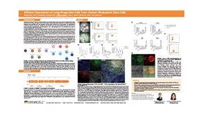 科学海报Efficient Generation of Lung Progenitor Cells From Human Pluripotent Stem Cells
 科学海报Single-Cell RNA Sequencing Analysis of Regionally Patterned Human Pluripotent Stem Cell-Derived Neural Organoids

 沪公网安备31010102008431号