Kleinstreuer NC et al. (NOV 2011)
Toxicology and Applied Pharmacology 257 1 111--121
Identifying developmental toxicity pathways for a subset of ToxCast chemicals using human embryonic stem cells and metabolomics
Metabolomics analysis was performed on the supernatant of human embryonic stem (hES) cell cultures exposed to a blinded subset of 11 chemicals selected from the chemical library of EPA's ToxCast™ chemical screening and prioritization research project. Metabolites from hES cultures were evaluated for known and novel signatures that may be indicative of developmental toxicity. Significant fold changes in endogenous metabolites were detected for 83 putatively annotated mass features in response to the subset of ToxCast chemicals. The annotations were mapped to specific human metabolic pathways. This revealed strong effects on pathways for nicotinate and nicotinamide metabolism,pantothenate and CoA biosynthesis,glutathione metabolism,and arginine and proline metabolism pathways. Predictivity for adverse outcomes in mammalian prenatal developmental toxicity studies used ToxRefDB and other sources of information,including Stemina Biomarker Discovery's predictive DevTox® model trained on 23 pharmaceutical agents of known developmental toxicity and differing potency. The model initially predicted developmental toxicity from the blinded ToxCast compounds in concordance with animal data with 73% accuracy. Retraining the model with data from the unblinded test compounds at one concentration level increased the predictive accuracy for the remaining concentrations to 83%. These preliminary results on a 11-chemical subset of the ToxCast chemical library indicate that metabolomics analysis of the hES secretome provides information valuable for predictive modeling and mechanistic understanding of mammalian developmental toxicity.
View Publication
产品号#:
05850
05857
05870
05875
85850
85857
85870
85875
产品名:
mTeSR™1
mTeSR™1
Busskamp V et al. (NOV 2014)
Molecular systems biology 10 11 760
Rapid neurogenesis through transcriptional activation in human stem cells.
Advances in cellular reprogramming and stem cell differentiation now enable ex vivo studies of human neuronal differentiation. However,it remains challenging to elucidate the underlying regulatory programs because differentiation protocols are laborious and often result in low neuron yields. Here,we overexpressed two Neurogenin transcription factors in human-induced pluripotent stem cells and obtained neurons with bipolar morphology in 4 days,at greater than 90% purity. The high purity enabled mRNA and microRNA expression profiling during neurogenesis,thus revealing the genetic programs involved in the rapid transition from stem cell to neuron. The resulting cells exhibited transcriptional,morphological and functional signatures of differentiated neurons,with greatest transcriptional similarity to prenatal human brain samples. Our analysis revealed a network of key transcription factors and microRNAs that promoted loss of pluripotency and rapid neurogenesis via progenitor states. Perturbations of key transcription factors affected homogeneity and phenotypic properties of the resulting neurons,suggesting that a systems-level view of the molecular biology of differentiation may guide subsequent manipulation of human stem cells to rapidly obtain diverse neuronal types.
View Publication
产品号#:
05854
05855
05850
05857
05870
05875
85850
85857
85870
85875
产品名:
mFreSR™
mFreSR™
mTeSR™1
mTeSR™1
Lungova V et al. ( 2014)
1307 237--243
Derivation of Epithelial Cells from Human Embryonic Stem Cells as an In Vitro Model of Vocal Mucosa
Vocal fold epithelial cells are very difficult to study as the vocal fold epithelial cell lines do not exist and they cannot be removed from the healthy larynx without engendering a significant and unacceptable risk to vocal fold function. Here,we describe the procedure to create an engineered vocal fold tissue construct consisting of the scaffold composed of the collagen 1 gel seeded with human fibroblasts and simple epithelial progenitors seeded on the scaffold and cultivated at air-liquid interface for 19-21 days to derive the stratified squamous epithelium. This model of vocal fold mucosa is very similar in morphology,gene expression,and phenotypic characteristics to native vocal fold epithelial cells and the underlying lamina propria and,therefore,offers a promising approach to studying vocal fold biology and biomechanics in health and disease.
View Publication
产品号#:
05850
05857
05870
05875
85850
85857
85870
85875
产品名:
mTeSR™1
mTeSR™1
Wang H-CC et al. (OCT 2014)
Cancer Informatics 13 Suppl 5 25--35
Profiling the microRNA Expression in Human iPS and iPS-derived Retinal Pigment Epithelium.
The purpose of this study is to characterize the microRNA (miRNA) expression profiles of induced pluripotent stem (iPS) cells and retinal pigment epithelium (RPE) derived from induced pluripotent stem cells (iPS-RPE). MiRNAs have been demonstrated to play critical roles in both maintaining pluripotency and facilitating differentiation. Gene expression networks accountable for maintenance and induction of pluripotency are linked and share components with those networks implicated in oncogenesis. Therefore,we hypothesize that miRNA expression profiling will distinguish iPS cells from their iPS-RPE progeny. To identify and analyze differentially expressed miRNAs,RPE was derived from iPS using a spontaneous differentiation method. MiRNA microarray analysis identified 155 probes that were statistically differentially expressed between iPS and iPS-RPE cells. Up-regulated miRNAs including miR-181c and miR-129-5p may play a role in promoting differentiation,while down-regulated miRNAs such as miR-367,miR-18b,and miR-20b are implicated in cell proliferation. Subsequent miRNA-target and network analysis revealed that these miRNAs are involved in cellular development,cell cycle progression,cell death,and survival. A systematic interrogation of temporal and spatial expression of iPS-RPE miRNAs and their associated target mRNAs will provide new insights into the molecular mechanisms of carcinogenesis,eye differentiation and development.
View Publication
产品号#:
05850
05857
05870
05875
85850
85857
85870
85875
产品名:
mTeSR™1
mTeSR™1
Hudson J et al. (JUN 2012)
Stem cells and development 21 9 1513--23
Primitive cardiac cells from human embryonic stem cells.
Pluripotent stem cell-derived cardiomyocytes are currently being investigated for in vitro human heart models and as potential therapeutics for heart failure. In this study,we have developed a differentiation protocol that minimizes the need for specific human embryonic stem cell (hESC) line optimization. We first reduced the heterogeneity that exists within the starting population of bulk cultured hESCs by using cells adapted to single-cell passaging in a 2-dimensional (2D) culture format. Compared with bulk cultures,single-cell cultures comprised larger fractions of TG30(hi)/OCT4(hi) cells,corresponding to an increased expression of pluripotency markers OCT4 and NANOG,and reduced expression of early lineage-specific markers. A 2D temporal differentiation protocol was then developed,aimed at reducing the inherent heterogeneity and variability of embryoid body-based protocols,with induction of primitive streak cells using bone morphogenetic protein 4 and activin A,followed by cardiogenesis via inhibition of Wnt signaling using the small molecules IWP-4 or IWR-1. IWP-4 treatment resulted in a large percentage of cells expressing low amounts of cardiac myosin heavy chain and expression of early cardiac progenitor markers ISL1 and NKX2-5,thus indicating the production of large numbers of immature cardiomyocytes (˜65,000/cm(2) or ˜1.5 per input hESC). This protocol was shown to be effective in HES3,H9,and,to a lesser,extent,MEL1 hESC lines. In addition,we observed that IWR-1 induced predominantly atrial myosin light chain (MLC2a) expression,whereas IWP-4 induced expression of both atrial (MLC2a) and ventricular (MLC2v) forms. The intrinsic flexibility and scalability of this 2D protocol mean that the output population of primitive cardiomyocytes will be particularly accessible and useful for the investigation of molecular mechanisms driving terminal cardiomyocyte differentiation,and potentially for the future treatment of heart failure.
View Publication
产品号#:
05850
05857
05870
05875
72552
72554
85850
85857
85870
85875
产品名:
IWP-4
IWP-4
mTeSR™1
mTeSR™1
Lu Y et al. (FEB 2012)
Stem cells and development 21 3 394--403
Avian-Induced Pluripotent Stem Cells Derived Using Human Reprogramming Factors
Avian species are important model animals for developmental biology and disease research. However,unlike in mice,where clonal lines of pluripotent stem cells have enabled researchers to study mammalian gene function,clonal and highly proliferative pluripotent avian cell lines have been an elusive goal. Here we demonstrate the generation of avian induced pluripotent stem cells (iPSCs),the first nonmammalian iPSCs,which were clonally isolated and propagated,important attributes not attained in embryo-sourced avian cells. This was accomplished using human pluripotency genes rather than avian genes,indicating that the process in which mammalian and nonmammalian cells are reprogrammed is a conserved process. Quail iPSCs (qiPSCs) were capable of forming all 3 germ layers in vitro and were directly differentiated in culture into astrocytes,oligodendrocytes,and neurons. Ultimately,qiPSCs were capable of generating live chimeric birds and incorporated into tissues from all 3 germ layers,extraembryonic tissues,and potentially the germline. These chimera competent qiPSCs and in vitro differentiated cells offer insight into the conserved nature of reprogramming and genetic tools that were only previously available in mammals.
View Publication
产品号#:
05850
05857
05870
05875
85850
85857
85870
85875
产品名:
mTeSR™1
mTeSR™1
Wu J et al. (APR 2015)
Stem cells and development 24 7 892--903
Increased culture density is linked to decelerated proliferation, prolonged G1 phase, and enhanced propensity for differentiation of self-renewing human pluripotent stem cells.
Human pluripotent stem cells (hPSCs) display a very short G1 phase and rapid proliferation kinetics. Regulation of the cell cycle,which is linked to pluripotency and differentiation,is dependent on the stem cell environment,particularly on culture density. This link has been so far empirical and central to disparities in the growth rates and fractions of self-renewing hPSCs residing in different cycle phases. In this study,hPSC cycle progression in conjunction with proliferation and differentiation were comprehensively investigated for different culture densities. Cell proliferation decelerated significantly at densities beyond 50×10(4) cells/cm(2). Correspondingly,the G1 fraction increased from 25% up to 60% at densities greater than 40×10(4) cells/cm(2) while still hPSC pluripotency marker expression was maintained. In parallel,expression of the cycle inhibitor CDKN1A (p21) was increased,while that of p27 and p53 did not change significantly. After 4 days of culture in an unconditioned medium,greater heterogeneity was noted in the differentiation outcomes and was limited by reducing the density variation. A quantitative model was constructed for self-renewing and differentiating hPSC ensembles to gain a better understanding of the link between culture density,cycle progression,and stem cell state. Results for multiple hPSC lines and medium types corroborated experimental findings. Media commonly used for maintenance of self-renewing hPSCs exhibited the slowest kinetics of induction of differentiation (kdiff),while BMP4 supplementation led to 14-fold higher kdiff values. Spontaneous differentiation in a growth factor-free medium exhibited the largest variation in outcomes at different densities. In conjunction with the quantitative framework,our findings will facilitate rationalizing the selection of cultivation conditions for the generation of stem cell therapeutics.
View Publication
产品号#:
05850
05857
05870
05875
05916
85850
85857
85870
85875
产品名:
TeSR™-E5
mTeSR™1
mTeSR™1
Miyazaki T and Suemori H ( 2015)
1235 97--104
Cryopreservation of human pluripotent stem cells: a general protocol.
Cryopreservation is an essential technique to preserve stem cells,semipermanently sustaining their potentials. There are two main approaches of cryopreservation for human pluripotent stem cells (hPSCs). The first is the vitrification,which involves instantaneous freeze and thaw of hPSCs. The second is the conventional slow-cooling method and a rapid thaw. Both cryopreservation protocols have been standardized and optimized to yield high survivability of hPSCs.
View Publication

 EasySep™小鼠TIL(CD45)正选试剂盒


 挂图Small Molecules, Big Impact in Pluripotent Stem Cell Research Overview of signaling pathways and small molecules in pluripotent stem cell research
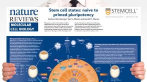 挂图Stem Cell States: Naive to Primed Pluripotency Properties of naive (ground) and primed pluripotent stem cells


 沪公网安备31010102008431号