Thomson JA et al. (NOV 1998)
Science (New York,N.Y.) 282 5391 1145--7
Embryonic stem cell lines derived from human blastocysts.
Human blastocyst-derived,pluripotent cell lines are described that have normal karyotypes,express high levels of telomerase activity,and express cell surface markers that characterize primate embryonic stem cells but do not characterize other early lineages. After undifferentiated proliferation in vitro for 4 to 5 months,these cells still maintained the developmental potential to form trophoblast and derivatives of all three embryonic germ layers,including gut epithelium (endoderm); cartilage,bone,smooth muscle,and striated muscle (mesoderm); and neural epithelium,embryonic ganglia,and stratified squamous epithelium (ectoderm). These cell lines should be useful in human developmental biology,drug discovery,and transplantation medicine.
View Publication
产品号#:
05860
05880
05850
05857
05870
05875
产品名:
Vittet D et al. (NOV 1996)
Blood 88 9 3424--31
Embryonic stem cells differentiate in vitro to endothelial cells through successive maturation steps.
The mechanisms involved in the regulation of vasculogenesis still remain unclear in mammals. Totipotent embryonic stem (ES) cells may represent a suitable in vitro model to study molecular events involved in vascular development. In this study,we followed the expression kinetics of a relatively large set of endothelial-specific markers in ES-derived embryoid bodies (EBs). Results of both reverse transcription-polymerase chain reaction and/or immunofluorescence analysis show that a spontaneous endothelial differentiation occurs during EBs development. ES-derived endothelial cells express a full range of cell lineage-specific markers: platelet endothelial cell adhesion molecule (PECAM),Flk-1,tie-1,tie-2,vascular endothelial (VE) cadherin,MECA-32,and MEC-14.7. Analysis of the kinetics of endothelial marker expression allows the distinction of successive maturation steps. Flk-1 was the first to be detected; its mRNA is apparent from day 3 of differentiation. PECAM and tie-2 mRNAs were found to be expressed only from day 4,whereas VE-cadherin and tie-1 mRNAs cannot be detected before day 5. Immunofluorescence stainings of EBs with antibodies directed against Flk-1,PECAM,VE-cadherin,MECA-32,and MEC-14.7 confirmed that the expression of these antigens occurs at different steps of endothelial cell differentiation. The addition of an angiogenic growth factor mixture including erythropoietin,interleukin-6,fibroblast growth factor 2,and vascular endothelial growth factor in the EB culture medium significantly increased the development of primitive vascular-like structures within EBs. These results indicate that this in vitro system contains a large part of the endothelial cell differentiation program and constitutes a suitable model to study the molecular mechanisms involved in vasculogenesis.
View Publication
产品号#:
06902
06952
00321
00322
00323
00324
00325
产品名:
Yamashita J et al. (NOV 2000)
Nature 408 6808 92--6
Flk1-positive cells derived from embryonic stem cells serve as vascular progenitors.
Interaction between endothelial cells and mural cells (pericytes and vascular smooth muscle) is essential for vascular development and maintenance. Endothelial cells arise from Flk1-expressing (Flk1+) mesoderm cells,whereas mural cells are believed to derive from mesoderm,neural crest or epicardial cells and migrate to form the vessel wall. Difficulty in preparing pure populations of these lineages has hampered dissection of the mechanisms underlying vascular formation. Here we show that Flk1+ cells derived from embryonic stem cells can differentiate into both endothelial and mural cells and can reproduce the vascular organization process. Vascular endothelial growth factor promotes endothelial cell differentiation,whereas mural cells are induced by platelet-derived growth factor-BB. Vascular cells derived from Flk1+ cells can organize into vessel-like structures consisting of endothelial tubes supported by mural cells in three-dimensional culture. Injection of Flk1+ cells into chick embryos showed that they can incorporate as endothelial and mural cells and contribute to the developing vasculature in vivo. Our findings indicate that Flk1+ cells can act as 'vascular progenitor cells' to form mature vessels and thus offer potential for tissue engineering of the vascular system.
View Publication
产品号#:
06902
06952
00321
00322
00323
00324
00325
产品名:
Lund RJ et al. (NOV 2013)
Stem Cell Research 11 3 1024--1036
Karyotypically abnormal human ESCs are sensitive to HDAC inhibitors and show altered regulation of genes linked to cancers and neurological diseases
Genomic abnormalities may accumulate in human embryonic stem cells (hESCs) during in vitro maintenance. Characterization of the mechanisms enabling survival and expansion of abnormal hESCs is important due to consequences of genetic changes for the therapeutic utilization of stem cells. Furthermore,these cells provide an excellent model to study transformation in vitro. We report here that the histone deacetylase proteins,HDAC1 and HDAC2,are increased in karyotypically abnormal hESCs when compared to their normal counterparts. Importantly,similar to many cancer cell lines,we found that HDAC inhibitors repress proliferation of the karyotypically abnormal hESCs,whereas normal cells are more resistant to the treatment. The decreased proliferation correlates with downregulation of HDAC1 and HDAC2 proteins,induction of the proliferation inhibitor,cyclin-dependent kinase inhibitor 1A (CDKN1A),and altered regulation of tumor suppressor protein Retinoblastoma 1 (RB1). Through genome-wide transcriptome analysis we have identified genes with altered expression and responsiveness to HDAC inhibition in abnormal cells. Most of these genes are linked to severe developmental and neurological diseases and cancers. Our results highlight the importance of epigenetic mechanisms in the regulation of genomic stability of hESCs,and provide valuable candidates for targeted and selective growth inhibition of karyotypically abnormal cells. textcopyright 2013 Elsevier B.V.
View Publication
产品号#:
05850
05857
05870
05875
85850
85857
85870
85875
产品名:
mTeSR™1
mTeSR™1
Wang J et al. (NOV 2013)
Biomaterials 34 35 8878--8886
Effect of engineered anisotropy on the susceptibility of human pluripotent stem cell-derived ventricular cardiomyocytes to arrhythmias
Human (h) pluripotent stem cells (PSC) such as embryonic stem cells (ESC) can be directed into cardiomyocytes (CMs),representing a potential unlimited cell source for disease modeling,cardiotoxicity screening and myocardial repair. Although the electrophysiology of single hESC-CMs is now better defined,their multi-cellular arrhythmogenicity has not been thoroughly assessed due to the lack of a suitable experimental platform. Indeed,the generation of ventricular (V) fibrillation requires single-cell triggers as well as sustained multi-cellular reentrant events. Although native VCMs are aligned in a highly organized fashion such that electrical conduction is anisotropic for coordinated contractions,hESC-derived CM (hESC-CM) clusters are heterogenous and randomly organized,and therefore not representative of native conditions. Here,we reported that engineered alignment of hESC-VCMs on biomimetic grooves uniquely led to physiologically relevant responses. Aligned but not isotropic control preparations showed distinct longitudinal (L) and transverse (T) conduction velocities (CV),resembling the native human V anisotropic ratio (AR=LCV/TCV=1.8-2.0). Importantly,the total incidence of spontaneous and inducible arrhythmias significantly reduced from 57% in controls to 17-23% of aligned preparations,thereby providing a physiological baseline for assessing arrhythmogenicity. As such,promotion of pro-arrhythmic effect (e.g.,spatial dispersion by ?? adrenergic stimulation) could be better predicted. Mechanistically,such anisotropy-induced electrical stability was not due to maturation of the cellular properties of hESC-VCMs but their physical arrangement. In conclusion,not only do functional anisotropic hESC-VCMs engineered by multi-scale topography represent a more accurate model for efficacious drug discovery and development as well as arrhythmogenicity screening (of pharmacological and genetic factors),but our approach may also lead to future transplantable prototypes with improved efficacy and safety against arrhythmias. ?? 2013.
View Publication

 EasySep™小鼠TIL(CD45)正选试剂盒


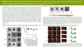 科学海报STEMdiff™ Cerebral Organoid Kit: A New Tool for the Culture of 3D Brain Organoids Derived from hPSCs

 沪公网安备31010102008431号