van der Meer AD et al. (SEP 2013)
Lab on a Chip 13 18 3562--3568
Three-dimensional co-cultures of human endothelial cells and embryonic stem cell-derived pericytes inside a microfluidic device
Organs-on-chips are microengineered in vitro tissue structures that can be used as platforms for physiological and pathological research. They provide tissue-like microenvironments in which different cell types can be co-cultured in a controlled manner to create synthetic organ mimics. Blood vessels are an integral part of all tissues in the human body. Development of vascular structures is therefore an important research topic for advancing the field of organs-on-chips since generated tissues will require a blood or nutrient supply. Here,we have engineered three-dimensional constructs of vascular tissue inside microchannels by injecting a mixture of human umbilical vein endothelial cells,human embryonic stem cell-derived pericytes (the precursors of vascular smooth muscle cells) and rat tail collagen I into a polydimethylsiloxane microfluidic channel with dimensions 500 μm × 120 μm × 1 cm (w × h × l). Over the course of 12 h,the cells organized themselves into a single long tube resembling a blood vessel that followed the contours of the channel. Detailed examination of tube morphology by confocal microscopy revealed a mature endothelial monolayer with complete PECAM-1 staining at cell–cell contacts and pericytes incorporated inside the tubular structures. We also demonstrated that tube formation was disrupted in the presence of a neutralizing antibody against transforming growth factor-beta (TGF-β). The TGF-β signaling pathway is essential for normal vascular development; deletion of any of its components in mouse development results in defective vasculogenesis and angiogenesis and mutations in humans have been linked to multiple vascular genetic diseases. In the engineered microvessels,inhibition of TGF-β signaling resulted in tubes with smaller diameters and higher tortuosity,highly reminiscent of the abnormal vessels observed in patients with one particular vascular disease known as hereditary hemorrhagic telangiectasia (HHT). In summary,we have developed microengineered three-dimensional vascular structures that can be used as a model to test the effects of drugs and study the interaction between different human vascular cell types. In the future,the model may be integrated into larger tissue constructs to advance the development of organs-on-chips.
View Publication
产品号#:
05850
05857
05870
05875
85850
85857
85870
85875
产品名:
mTeSR™1
mTeSR™1
Zhang Y et al. (JUN 2013)
Neuron 78 5 785--798
Rapid single-step induction of functional neurons from human pluripotent stem cells
Available methods for differentiating human embryonic stem cells (ESCs) and induced pluripotent cells (iPSCs) into neurons are often cumbersome,slow,and variable. Alternatively,human fibroblasts can be directly converted into induced neuronal (iN) cells. However,with present techniques conversion is inefficient,synapse formation is limited,and only small amounts of neurons can be generated. Here,we show that human ESCs and iPSCs can be converted into functional iN cells with nearly 100% yield and purity in less than 2weeks by forced expression of a single transcription factor. The resulting ES-iN or iPS-iN cells exhibit quantitatively reproducible properties independent of the cell line of origin,form mature pre- and postsynaptic specializations,and integrate into existing synaptic networks when transplanted into mouse brain. As illustrated by selected examples,our approach enables large-scale studies of human neurons for questions such as analyses of human diseases,examination of human-specific genes,and drug screening
View Publication
Kreitzer FR et al. (JUN 2013)
American journal of stem cells 2 2 119--31
A robust method to derive functional neural crest cells from human pluripotent stem cells.
Neural crest (NC) cells contribute to the development of many complex tissues of all three germ layers during embryogenesis,and its abnormal development accounts for several congenital birth defects. Generating NC cells-including specific subpopulations such as cranial,cardiac,and trunk NC cells-from human pluripotent stem cells will provide a valuable model system to study human development and disease. Here,we describe a rapid and robust NC differentiation method called LSB-short" that is based on dual SMAD pathway inhibition. This protocol yields high percentages of NC cell populations from multiple human induced pluripotent stem and human embryonic stem cell lines in 8 days. The resulting cells can be propagated easily�
View Publication
Fraga AM et al. (MAR 2011)
Cell Transplantation 20 3 431--40
Establishment of a Brazilian line of human embryonic stem cells in defined medium: implications for cell therapy in an ethnically diverse population.
Pluripotent human embryonic stem (hES) cells are an important experimental tool for basic and applied research,and a potential source of different tissues for transplantation. However,one important challenge for the clinical use of these cells is the issue of immunocompatibility,which may be dealt with by the establishment of hES cell banks to attend different populations. Here we describe the derivation and characterization of a line of hES cells from the Brazilian population,named BR-1,in commercial defined medium. In contrast to the other hES cell lines established in defined medium,BR-1 maintained a stable normal karyotype as determined by genomic array analysis after 6 months in continuous culture (passage 29). To our knowledge,this is the first reported line of hES cells derived in South America. We have determined its genomic ancestry and compared the HLA-profile of BR-1 and another 22 hES cell lines established elsewhere with those of the Brazilian population,finding they would match only 0.011% of those individuals. Our results highlight the challenges involved in hES cell banking for populations with a high degree of ethnic admixture.
View Publication
产品号#:
05850
05857
05870
05875
85850
85857
85870
85875
产品名:
mTeSR™1
mTeSR™1
Leung HW et al. (FEB 2011)
Tissue engineering. Part C,Methods 17 2 165--72
Agitation can induce differentiation of human pluripotent stem cells in microcarrier cultures.
One of the factors that can impact human embryonic stem cell expansion in stirred microcarrier culture reactors is mechanical stress caused by agitation. Therefore,we have investigated the effects of agitation on human embryonic stem cell growth and expression of pluripotent markers. Agitation of HES-2 cell line in microcarrier cultures in stirred spinner and agitated six-well plates did not affect expression of pluripotent markers,cell viability,and cell doubling times even after seven passages. However,HES-3 cell line was found to be shear sensitive,showing downregulation of three pluripotent markers Oct-4,mAb 84,and Tra-1-60,and lower cell densities in agitated as compared with static cultures,even after one passage. Cell viability was unaffected. The HES-3-agitated cultures showed increased expression of genes and proteins of the three germ layers. We were unable to prevent loss of pluripotent markers or restore doubling times in agitated HES-3 microcarrier cultures by addition of five different known cell protective polymers. In addition,the human induced pluripotent cell line IMR90 was also shown to differentiate in agitated conditions. These results indicate that the effect of agitation on cell growth and differentiation is cell line specific. We assume that the changes in the growth and differentiation of the agitation-sensitive (HES-3) cell line do not result from the effect of shear stress directly on cell viability,but rather by signaling effects that influence the cells to differentiate resulting in slower growth.
View Publication
产品号#:
05850
05857
05870
05875
85850
85857
85870
85875
产品名:
mTeSR™1
mTeSR™1
Calvanese V et al. (AUG 2010)
Proceedings of the National Academy of Sciences of the United States of America 107 31 13736--41
Sirtuin 1 regulation of developmental genes during differentiation of stem cells
The longevity-promoting NAD+-dependent class III histone deacetylase Sirtuin 1 (SIRT1) is involved in stem cell function by controlling cell fate decision and/or by regulating the p53-dependent expression of NANOG. We show that SIRT1 is down-regulated precisely during human embryonic stem cell differentiation at both mRNA and protein levels and that the decrease in Sirt1 mRNA is mediated by a molecular pathway that involves the RNA-binding protein HuR and the arginine methyltransferase coactivator-associated arginine methyltransferase 1 (CARM1). SIRT1 down-regulation leads to reactivation of key developmental genes such as the neuroretinal morphogenesis effectors DLL4,TBX3,and PAX6,which are epigenetically repressed by this histone deacetylase in pluripotent human embryonic stem cells. Our results indicate that SIRT1 is regulated during stem cell differentiation in the context of a yet-unknown epigenetic pathway that controls specific developmental genes in embryonic stem cells.
View Publication
产品号#:
05850
05857
05870
05875
85850
85857
85870
85875
产品名:
mTeSR™1
mTeSR™1
Bratt-Leal A et al. (JAN 2011)
Biomaterials 32 1 48--56
Incorporation of biomaterials in multicellular aggregates modulates pluripotent stem cell differentiation.
Biomaterials are increasingly being used to engineer the biochemical and biophysical properties of the extracellular stem cell microenvironment in order to tailor niche characteristics and direct cell phenotype. To date,stem cell-biomaterial interactions have largely been studied by introducing stem cells into artificial environments,such as 2D cell culture on biomaterial surfaces,encapsulation of cell suspensions within hydrogel materials,or cell seeding on 3D polymeric scaffolds. In this study,microparticles fabricated from different materials,such as agarose,PLGA and gelatin,were stably integrated,in a dose-dependent manner,within aggregates of pluripotent stem cells (PSCs) prior to differentiation as a means to directly examine stem cell-biomaterial interactions in 3D. Interestingly,the presence of the materials within the stem cell aggregates differentially modulated the gene and protein expression patterns of several differentiation markers without adversely affecting cell viability. Microparticle incorporation within 3D stem cell aggregates can control the spatial presentation of extracellular environmental cues (i.e. soluble factors,extracellular matrix and intercellular adhesion molecules) as a means to direct the differentiation of stem cells for tissue engineering and regenerative medicine applications. In addition,these results suggest that the physical presence of microparticles within stem cell aggregates does not compromise PSC differentiation,but in fact the choice of biomaterials can impact the propensity of stem cells to adopt particular differentiated cell phenotypes.
View Publication

 EasySep™小鼠TIL(CD45)正选试剂盒


 科学海报Single-Cell RNA Sequencing Analysis of Regionally Patterned Human Pluripotent Stem Cell-Derived Neural Organoids
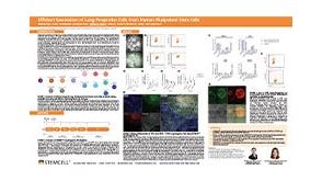 科学海报Efficient Generation of Lung Progenitor Cells From Human Pluripotent Stem Cells


 沪公网安备31010102008431号