Induced pluripotent stem cells with a mitochondrial dna deletion
In congenital mitochondrial DNA (mtDNA) disorders,a mixture of normal and mutated mtDNA (termed heteroplasmy) exists at varying levels in different tissues,which determines the severity and phenotypic expression of disease. Pearson marrow pancreas syndrome (PS) is a congenital bone marrow failure disorder caused by heteroplasmic deletions in mtDNA. The cause of the hematopoietic failure in PS is unknown,and adequate cellular and animal models are lacking. Induced pluripotent stem (iPS) cells are particularly amenable for studying mtDNA disorders,as cytoplasmic genetic material is retained during direct reprogramming. Here,we derive and characterize iPS cells from a patient with PS. Taking advantage of the tendency for heteroplasmy to change with cell passage,we isolated isogenic PS-iPS cells without detectable levels of deleted mtDNA. We found that PS-iPS cells carrying a high burden of deleted mtDNA displayed differences in growth,mitochondrial function,and hematopoietic phenotype when differentiated in vitro,compared to isogenic iPS cells without deleted mtDNA. Our results demonstrate that reprogramming somatic cells from patients with mtDNA disorders can yield pluripotent stem cells with varying burdens of heteroplasmy that might be useful in the study and treatment of mitochondrial diseases. STEM CELLS2013;31:1287–1297
View Publication
Kreitzer FR et al. (JUN 2013)
American journal of stem cells 2 2 119--31
A robust method to derive functional neural crest cells from human pluripotent stem cells.
Neural crest (NC) cells contribute to the development of many complex tissues of all three germ layers during embryogenesis,and its abnormal development accounts for several congenital birth defects. Generating NC cells-including specific subpopulations such as cranial,cardiac,and trunk NC cells-from human pluripotent stem cells will provide a valuable model system to study human development and disease. Here,we describe a rapid and robust NC differentiation method called LSB-short" that is based on dual SMAD pathway inhibition. This protocol yields high percentages of NC cell populations from multiple human induced pluripotent stem and human embryonic stem cell lines in 8 days. The resulting cells can be propagated easily�
View Publication
Rapid single-step induction of functional neurons from human pluripotent stem cells
Available methods for differentiating human embryonic stem cells (ESCs) and induced pluripotent cells (iPSCs) into neurons are often cumbersome,slow,and variable. Alternatively,human fibroblasts can be directly converted into induced neuronal (iN) cells. However,with present techniques conversion is inefficient,synapse formation is limited,and only small amounts of neurons can be generated. Here,we show that human ESCs and iPSCs can be converted into functional iN cells with nearly 100% yield and purity in less than 2weeks by forced expression of a single transcription factor. The resulting ES-iN or iPS-iN cells exhibit quantitatively reproducible properties independent of the cell line of origin,form mature pre- and postsynaptic specializations,and integrate into existing synaptic networks when transplanted into mouse brain. As illustrated by selected examples,our approach enables large-scale studies of human neurons for questions such as analyses of human diseases,examination of human-specific genes,and drug screening
View Publication
产品号#:
05850
05857
05870
05875
85850
85857
85870
85875
产品名:
mTeSR™1
mTeSR™1
van der Meer AD et al. (SEP 2013)
Lab on a Chip 13 18 3562--3568
Three-dimensional co-cultures of human endothelial cells and embryonic stem cell-derived pericytes inside a microfluidic device
Organs-on-chips are microengineered in vitro tissue structures that can be used as platforms for physiological and pathological research. They provide tissue-like microenvironments in which different cell types can be co-cultured in a controlled manner to create synthetic organ mimics. Blood vessels are an integral part of all tissues in the human body. Development of vascular structures is therefore an important research topic for advancing the field of organs-on-chips since generated tissues will require a blood or nutrient supply. Here,we have engineered three-dimensional constructs of vascular tissue inside microchannels by injecting a mixture of human umbilical vein endothelial cells,human embryonic stem cell-derived pericytes (the precursors of vascular smooth muscle cells) and rat tail collagen I into a polydimethylsiloxane microfluidic channel with dimensions 500 μm × 120 μm × 1 cm (w × h × l). Over the course of 12 h,the cells organized themselves into a single long tube resembling a blood vessel that followed the contours of the channel. Detailed examination of tube morphology by confocal microscopy revealed a mature endothelial monolayer with complete PECAM-1 staining at cell–cell contacts and pericytes incorporated inside the tubular structures. We also demonstrated that tube formation was disrupted in the presence of a neutralizing antibody against transforming growth factor-beta (TGF-β). The TGF-β signaling pathway is essential for normal vascular development; deletion of any of its components in mouse development results in defective vasculogenesis and angiogenesis and mutations in humans have been linked to multiple vascular genetic diseases. In the engineered microvessels,inhibition of TGF-β signaling resulted in tubes with smaller diameters and higher tortuosity,highly reminiscent of the abnormal vessels observed in patients with one particular vascular disease known as hereditary hemorrhagic telangiectasia (HHT). In summary,we have developed microengineered three-dimensional vascular structures that can be used as a model to test the effects of drugs and study the interaction between different human vascular cell types. In the future,the model may be integrated into larger tissue constructs to advance the development of organs-on-chips.
View Publication
产品号#:
05850
05857
05870
05875
85850
85857
85870
85875
产品名:
mTeSR™1
mTeSR™1
Kumagai H et al. (MAY 2013)
Biochemical and Biophysical Research Communications 434 4 710--716
Identification of small molecules that promote human embryonic stem cell self-renewal
Human embryonic stem cells (hESCs) and induced pluripotent cells have the potential to provide an unlimited source of tissues for regenerative medicine. For this purpose,development of defined/xeno-free culture systems under feeder-free conditions is essential for the expansion of hESCs. Most defined/xeno-free media for the culture of hESCs contain basic fibroblast growth factor (bFGF). Therefore,bFGF is thought to have an almost essential role for the expansion of hESCs in an undifferentiated state. Here,we report identification of small molecules,some of which were neurotransmitter antagonists (trimipramine and ethopropazine),which promote long-term hESC self-renewal without bFGF in the medium. The hESCs maintained high expression levels of pluripotency markers,had a normal karyotype after 20 passages,and could differentiate into all three germ layers. ?? 2013 Elsevier Inc.
View Publication

 EasySep™小鼠TIL(CD45)正选试剂盒


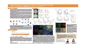 科学海报Efficient Generation of Lung Progenitor Cells From Human Pluripotent Stem Cells


 沪公网安备31010102008431号