Ji M et al. (SEP 2013)
Science Translational Medicine 5 201 201ra119--201ra119
Rapid, Label-Free Detection of Brain Tumors with Stimulated Raman Scattering Microscopy
Surgery is an essential component in the treatment of brain tumors. However,delineating tumor from normal brain remains a major challenge. We describe the use of stimulated Raman scattering (SRS) microscopy for differentiating healthy human and mouse brain tissue from tumor-infiltrated brain based on histoarchitectural and biochemical differences. Unlike traditional histopathology,SRS is a label-free technique that can be rapidly performed in situ. SRS microscopy was able to differentiate tumor from nonneoplastic tissue in an infiltrative human glioblastoma xenograft mouse model based on their different Raman spectra. We further demonstrated a correlation between SRS and hematoxylin and eosin microscopy for detection of glioma infiltration (κ = 0.98). Finally,we applied SRS microscopy in vivo in mice during surgery to reveal tumor margins that were undetectable under standard operative conditions. By providing rapid intraoperative assessment of brain tissue,SRS microscopy may ultimately improve the safety and accuracy of surgeries where tumor boundaries are visually indistinct.
View Publication
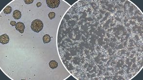
Kim MY et al. (MAR 2017)
Oncology letters 13 3 1767--1774
Accumulation of low-dose BIX01294 promotes metastatic potential of U251 glioblastoma cells.
BIX01294 (Bix) is known to be a euchromatic histone-lysine N-methyltransferase 2 inhibitor and treatment with Bix suppresses cancer cell survival and proliferation. In the present study,it was observed that sequential treatment with low-dose Bix notably increases glioblastoma cell migration and metastasis. It was demonstrated that U251 cells sequentially treated with low-dose Bix exhibited induced characteristic changes in critical epithelial-mesenchymal transition (EMT) markers,including E-cadherin,N-cadherin,β-catenin and zinc finger protein SNAI2. Notably,sequential treatment with Bix also increased the expression of cancer stem cell-associated markers,including sex determining region Y-box 2,octamer-binding transcription factor 4 and cluster of differentiation 133. Neurosphere formation was significantly enhanced in cells sequentially treated with Bix,compared with control cells (control: P=0.011; single treatment of Bix,P=0.045). The results of the present study suggest that accumulation of low-dose Bix enhanced the migration and metastatic potential of glioblastoma cells by regulating EMT-associated gene expression,which may be the cause of the altered properties of glioblastoma stem cells.
View Publication

 EasySep™小鼠TIL(CD45)正选试剂盒


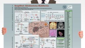 挂图SnapShot: Glioblastoma Multiforme Overview of the key concepts and mechanisms in glioblastoma multiforme biology
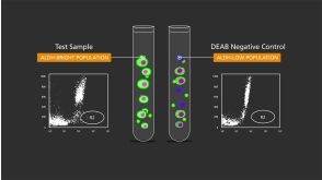
 技术公告Identification of Viable Stem and Progenitor Cells with ALDEFLUOR™


 沪公网安备31010102008431号