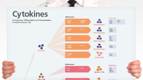
Human Immune Cytokines
Infographic of key cytokines for expansion, differentiation and characterization of major immune cell types
Chevalier MF et al. ( 2015)
The Journal of Infectious Diseases 211 5 769--779
Phenotype Alterations in Regulatory T-Cell Subsets in Primary HIV Infection and Identification of Tr1-like Cells as the Main Interleukin 10-Producing CD4+ T Cells
BACKGROUND: Conventional regulatory T cells (Tregs) can suppress human immunodeficiency virus type 1 (HIV-1)-specific immune responses but cannot control immune activation in primary HIV infection. Here,we characterized Treg subsets,using recently defined phenotypic delineation,and analyzed the relative contribution of cell subsets to the production of immunosuppressive cytokines in primary HIV infection. METHODS: In a longitudinal prospective study,ex vivo phenotyping of fresh peripheral blood mononuclear cells from patients with primary HIV infection was performed at baseline and month 6 of follow-up to characterize Treg subsets,immune activation,and cytokine production in isolated CD4(+) T cells. RESULTS: The frequency of CD4(+)CD25(+)CD127(low) Tregs and the distribution between the naive,memory,and activated/memory Treg subsets was similar in patients and healthy donors. However,Tregs from patients with primary HIV infection showed peculiar phenotypic profiles,such as elevated FoxP3,ICOS,and CTLA-4 expression,with CTLA-4 expression strikingly increased in all Treg subsets both at baseline and month 6 of follow-up. The great majority of interleukin 10 (IL-10)-producing CD4(+) T cells were FoxP3(neg) (ie,Tr1-like cells). In contrast to conventional Tregs,Tr1-like cells were inversely correlated with immune activation and not associated with lower effector T-cell responses. CONCLUSION: FoxP3(neg) Tr1-like cells-major contributors to IL-10 production-may have a beneficial role by controlling immune activation in early HIV infection.
View Publication
产品号#:
18062
18062RF
15022
15062
18251
18251RF
21000
20119
20155
15021
15061
产品名:
RosetteSep™人CD4+ T细胞富集抗体混合物
RosetteSep™人CD4+ T细胞富集抗体混合物
RoboSep™- S
RoboSep™ 吸头组件抛光剂
RoboSep™分选管套装(9个塑料管)
RosetteSep™人T细胞富集抗体混合物
RosetteSep™人T细胞富集抗体混合物
S. Bhatia et al. (may 2019)
Cancer research 79 10 2722--2735
Inhibition of EphB4-Ephrin-B2 Signaling Reprograms the Tumor Immune Microenvironment in Head and Neck Cancers.
Identifying targets present in the tumor microenvironment that contribute to immune evasion has become an important area of research. In this study,we identified EphB4-ephrin-B2 signaling as a regulator of both innate and adaptive components of the immune system. EphB4 belongs to receptor tyrosine kinase family that interacts with ephrin-B2 ligand at sites of cell-cell contact,resulting in bidirectional signaling. We found that EphB4-ephrin-B2 inhibition alone or in combination with radiation (RT) reduced intratumoral regulatory T cells (Tregs) and increased activation of both CD8+ and CD4+Foxp3- T cells compared with the control group in an orthotopic head and neck squamous cell carcinoma (HNSCC) model. We also compared the effect of EphB4-ephrin-B2 inhibition combined with RT with combined anti-PDL1 and RT and observed similar tumor growth suppression,particularly at early time-points. A patient-derived xenograft model showed reduction of tumor-associated M2 macrophages and favored polarization towards an antitumoral M1 phenotype following EphB4-ephrin-B2 inhibition with RT. In vitro,EphB4 signaling inhibition decreased Ki67-expressing Tregs and Treg activation compared with the control group. Overall,our study is the first to implicate the role of EphB4-ephrin-B2 in tumor immune response. Moreover,our findings suggest that EphB4-ephrin-B2 inhibition combined with RT represents a potential alternative for patients with HNSCC and could be particularly beneficial for patients who are ineligible to receive or cannot tolerate anti-PDL1 therapy. SIGNIFICANCE: These findings present EphB4-ephrin-B2 inhibition as an alternative to anti-PDL1 therapeutics that can be used in combination with radiation to induce an effective antitumor immune response in patients with HNSCC.
View Publication
Antunes I et al. (DEC 2010)
Journal of virology 84 24 12564--75
Suppression of innate immune pathology by regulatory T cells during Influenza A virus infection of immunodeficient mice.
The viral infection of higher vertebrates elicits potent innate and adaptive host immunity. However,an excessive or inappropriate immune response also may lead to host pathology that often is more severe than the direct effects of viral replication. Therefore,several mechanisms exist that regulate the magnitude and class of the immune response. Here,we have examined the potential involvement of regulatory T (Treg) cells in limiting pathology induced by influenza A virus (IAV) infection. Using lymphocyte-deficient mice as hosts,we showed that Treg cell reconstitution resulted in a significant delay in weight loss and prolonged survival following infection. The adoptively transferred Treg cells did not affect the high rate of IAV replication in the lungs of lymphocyte-deficient hosts,and therefore their disease-ameliorating effect was mediated through the suppression of innate immune pathology. Mechanistically,Treg cells reduced the accumulation and altered the distribution of monocytes/macrophages in the lungs of IAV-infected hosts. This reduction in lung monocytosis was associated with a specific delay in monocyte chemotactic protein-2 (MCP-2) induction in the infected lungs. Nevertheless,Treg cells failed to prevent the eventual development of severe disease in lymphocyte-deficient hosts,which likely was caused by the ongoing IAV replication. Indeed,using T-cell-deficient mice,which mounted a T-cell-independent B cell response to IAV,we further showed that the combination of virus-neutralizing antibodies and transferred Treg cells led to the complete prevention of clinical disease following IAV infection. Taken together,these results suggested that innate immune pathology and virus-induced pathology are the two main contributors to pathogenesis during IAV infection.
View Publication
产品号#:
19782
19792
产品名:
Feng T et al. (NOV 2010)
Journal of immunology (Baltimore,Md. : 1950) 185 10 5915--25
Generation of mucosal dendritic cells from bone marrow reveals a critical role of retinoic acid.
It is unknown how dendritic cells (DCs) become specialized as mucosal DCs and maintain intestinal homeostasis. We report that a subset of bone marrow cells freshly isolated from C57BL/6 mice express the retinoic acid (RA)-synthesizing enzyme aldehyde dehydrogenase family 1,subfamily A2 (ALDH1a2) and are capable of providing RA to DC precursors in the bone marrow microenvironment. RA induced bone marrow-derived DCs to express CCR9 and ALDH1a2 and conferred upon them mucosal DC functions,including induction of Foxp3(+) regulatory T cells,IgA-secreting B cells,and gut-homing molecules. This response of DCs to RA was dependent on a narrow time window and stringent dose effect. RA promoted bone marrow-derived DC production of bioactive TGF-β by inhibiting suppressor of cytokine signaling 3 expression and thereby enhancing STAT3 activation. These RA effects were evident in vivo,in that mucosal DCs from vitamin A-deficient mice had reduced mucosal DC function,namely failure to induce Foxp3(+) regulatory T cells. Furthermore,MyD88 signaling enhanced RA-educated DC ALDH1a2 expression and was required for optimal TGF-β production. These data indicate that RA plays a critical role in the generation of mucosal DCs from bone marrow and in their functional activity.
View Publication

 EasySep™小鼠TIL(CD45)正选试剂盒


 挂图Human Immune Cytokines Infographic of key cytokines for expansion, differentiation and characterization of major immune cell types

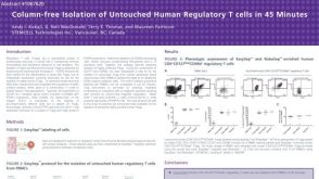 科学海报Immunomagnetic Cell Isolation of Untouched Human Regulatory T Cells

 挂图Frequencies of Immune Cells in Rat Tissue Lists the estimated frequencies of more than 15 immune cell types in Sprague Dawley rats

 沪公网安备31010102008431号