Khazen R et al. (MAR 2016)
Nature Communications 7 10823
Melanoma cell lysosome secretory burst neutralizes the CTL-mediated cytotoxicity at the lytic synapse.
Human melanoma cells express various tumour antigens that are recognized by CD8(+) cytotoxic T lymphocytes (CTLs) and elicit tumour-specific responses in vivo. However,natural and therapeutically enhanced CTL responses in melanoma patients are of limited efficacy. The mechanisms underlying CTL effector phase failure when facing melanomas are still largely elusive. Here we show that,on conjugation with CTL,human melanoma cells undergo an active late endosome/lysosome trafficking,which is intensified at the lytic synapse and is paralleled by cathepsin-mediated perforin degradation and deficient granzyme B penetration. Abortion of SNAP-23-dependent lysosomal trafficking,pH perturbation or impairment of lysosomal proteolytic activity restores susceptibility to CTL attack. Inside the arsenal of melanoma cell strategies to escape immune surveillance,we identify a self-defence mechanism based on exacerbated lysosome secretion and perforin degradation at the lytic synapse. Interfering with this synaptic self-defence mechanism might be useful in potentiating CTL-mediated therapies in melanoma patients.
View Publication
产品号#:
17953
17953RF
100-0710
产品名:
EasySep™人CD8+ T细胞分选试剂盒
RoboSep™ 人CD8+ T细胞分选试剂盒
EasySep™人CD8+ T细胞分选试剂盒
Thompson EA et al. (APR 2016)
Journal of Immunology 196 7 3054--63
Shortened Intervals during Heterologous Boosting Preserve Memory CD8 T Cell Function but Compromise Longevity.
Developing vaccine strategies to generate high numbers of Ag-specific CD8 T cells may be necessary for protection against recalcitrant pathogens. Heterologous prime-boost-boost immunization has been shown to result in large quantities of functional memory CD8 T cells with protective capacities and long-term stability. Completing the serial immunization steps for heterologous prime-boost-boost can be lengthy,leaving the host vulnerable for an extensive period of time during the vaccination process. We show in this study that shortening the intervals between boosting events to 2 wk results in high numbers of functional and protective Ag-specific CD8 T cells. This protection is comparable to that achieved with long-term boosting intervals. Short-boosted Ag-specific CD8 T cells display a canonical memory T cell signature associated with long-lived memory and have identical proliferative potential to long-boosted T cells Both populations robustly respond to antigenic re-exposure. Despite this,short-boosted Ag-specific CD8 T cells continue to contract gradually over time,which correlates to metabolic differences between short- and long-boosted CD8 T cells at early memory time points. Our studies indicate that shortening the interval between boosts can yield abundant,functional Ag-specific CD8 T cells that are poised for immediate protection; however,this is at the expense of forming stable long-term memory.
View Publication
产品号#:
17951
17951RF
17952
17952RF
19254
19254RF
19853
19853RF
100-0695
100-0696
产品名:
EasySep™人T细胞分选试剂盒
RoboSep™ 人T细胞分选试剂盒
EasySep™人CD4+ T细胞分选试剂盒
RoboSep™ 人CD4+ T细胞分选试剂盒
EasySep™人Naïve B细胞富集试剂盒
RoboSep™ 人Naïve B细胞富集试剂盒含滤芯吸头
EasySep™小鼠CD8+ T细胞分选试剂盒
RoboSep™ 小鼠CD8+ T细胞分选试剂盒
EasySep™人T细胞分选试剂盒
EasySep™人CD4+ T细胞分离试剂盒
Deets KA et al. (MAR 2016)
Journal of Immunology 196 6 2450--5
Cutting Edge: Enhanced Clonal Burst Size Corrects an Otherwise Defective Memory Response by CD8+ Recent Thymic Emigrants.
The youngest peripheral T cells (recent thymic emigrants [RTEs]) are functionally distinct from naive T cells that have completed postthymic maturation. We assessed the RTE memory response and found that RTEs produced less granzyme B than their mature counterparts during infection but proliferated more and,therefore,generated equivalent target killing in vivo. Postinfection,RTE numbers contracted less dramatically than those of mature T cells,but RTEs were delayed in their transition to central memory,displaying impaired expression of CD62L,IL-2,Eomesodermin,and CXCR4,which resulted in impaired bone marrow localization. RTE-derived and mature memory cells expanded equivalently during rechallenge,indicating that the robust proliferative capacity of RTEs was maintained independently of central memory phenotype. Thus,the diminished effector function and delayed central memory differentiation of RTE-derived memory cells are counterbalanced by their increased proliferative capacity,driving the efficacy of the RTE response to that of mature T cells.
View Publication
产品号#:
19853
19853RF
产品名:
EasySep™小鼠CD8+ T细胞分选试剂盒
RoboSep™ 小鼠CD8+ T细胞分选试剂盒
Gracias DT et al. (FEB 2016)
Journal of Immunology 196 3 1186--98
Phosphatidylinositol 3-Kinase p110δ Isoform Regulates CD8+ T Cell Responses during Acute Viral and Intracellular Bacterial Infections.
The p110δ isoform of PI3K is known to play an important role in immunity,yet its contribution to CTL responses has not been fully elucidated. Using murine p110δ-deficient CD8(+) T cells,we demonstrated a critical role for the p110δ subunit in the generation of optimal primary and memory CD8(+) T cell responses. This was demonstrated in both acute viral and intracellular bacterial infections in mice. We show that p110δ signaling is required for CD8(+) T cell activation,proliferation and effector cytokine production. We provide evidence that the effects of p110δ signaling are mediated via Akt activation and through the regulation of TCR-activated oxidative phosphorylation and aerobic glycolysis. In light of recent clinical trials that employ drugs targeting p110δ in certain cancers and other diseases,our study suggests caution in using these drugs in patients,as they could potentially increase susceptibility to infectious diseases. These studies therefore reveal a novel and direct role for p110δ signaling in in vivo CD8(+) T cell immunity to microbial pathogens.
View Publication
产品号#:
19858
19858RF
产品名:
EasySep™小鼠Naïve CD8+ T细胞分选试剂盒
RoboSep™ 小鼠Naïve CD8+ T细胞分选试剂盒
Chevalier MF et al. ( 2015)
The Journal of Infectious Diseases 211 5 769--779
Phenotype Alterations in Regulatory T-Cell Subsets in Primary HIV Infection and Identification of Tr1-like Cells as the Main Interleukin 10-Producing CD4+ T Cells
BACKGROUND: Conventional regulatory T cells (Tregs) can suppress human immunodeficiency virus type 1 (HIV-1)-specific immune responses but cannot control immune activation in primary HIV infection. Here,we characterized Treg subsets,using recently defined phenotypic delineation,and analyzed the relative contribution of cell subsets to the production of immunosuppressive cytokines in primary HIV infection. METHODS: In a longitudinal prospective study,ex vivo phenotyping of fresh peripheral blood mononuclear cells from patients with primary HIV infection was performed at baseline and month 6 of follow-up to characterize Treg subsets,immune activation,and cytokine production in isolated CD4(+) T cells. RESULTS: The frequency of CD4(+)CD25(+)CD127(low) Tregs and the distribution between the naive,memory,and activated/memory Treg subsets was similar in patients and healthy donors. However,Tregs from patients with primary HIV infection showed peculiar phenotypic profiles,such as elevated FoxP3,ICOS,and CTLA-4 expression,with CTLA-4 expression strikingly increased in all Treg subsets both at baseline and month 6 of follow-up. The great majority of interleukin 10 (IL-10)-producing CD4(+) T cells were FoxP3(neg) (ie,Tr1-like cells). In contrast to conventional Tregs,Tr1-like cells were inversely correlated with immune activation and not associated with lower effector T-cell responses. CONCLUSION: FoxP3(neg) Tr1-like cells-major contributors to IL-10 production-may have a beneficial role by controlling immune activation in early HIV infection.
View Publication
Human Immune Cytokines
Infographic of key cytokines for expansion, differentiation and characterization of major immune cell types
Hassanzadeh-Kiabi N et al. (NOV 2016)
Journal of immunology (Baltimore,Md. : 1950)
Autocrine Type I IFN Signaling in Dendritic Cells Stimulated with Fungal β-Glucans or Lipopolysaccharide Promotes CD8 T Cell Activation.
Type I IFNs are key mediators of immune defense against viruses and bacteria. Type I IFNs were also previously implicated in protection against fungal infection,but their roles in antifungal immunity have not been thoroughly investigated. A recent study demonstrated that bacterial and fungal β-glucans stimulate IFN-β production by dendritic cells (DCs) following detection by the Dectin-1 receptor,but the effects of β-glucan-induced type I IFNs have not been defined. We investigated whether type I IFNs regulate CD8 T cell activation by fungal β-glucan particle-stimulated DCs. We demonstrate that β-glucan-stimulated DCs induce CD8 T cell proliferation,activation marker (CD44 and CD69) expression,and production of IFN-γ,IL-2,and granzyme B. Moreover,we show that type I IFNs support robust CD8 T cell activation (proliferation and IFN-γ and granzyme B production) by β-glucan-stimulated DCs in vitro and in vivo due to autocrine effects on the DCs. Specifically,type I IFNs promote Ag presentation on MHC I molecules,CD86 and CD40 expression,and the production of IL-12 p70,IL-2,IL-6,and TNF-α by β-glucan-stimulated DCs. We also demonstrate a role for autocrine type I IFN signaling in bacterial LPS-induced DC maturation,although,in the context of LPS stimulation,this mechanism is not so critical for CD8 T cell activation (promotes IFN-γ production but not proliferation or granzyme B production). This study provides insight into the mechanisms underlying CD8 T cell activation during infection,which may be useful in the rational design of vaccines directed against pathogens and tumors.
View Publication
产品号#:
19858
19858RF
产品名:
EasySep™小鼠Naïve CD8+ T细胞分选试剂盒
RoboSep™ 小鼠Naïve CD8+ T细胞分选试剂盒
Ozga AJ et al. (OCT 2016)
The Journal of experimental medicine
pMHC affinity controls duration of CD8+ T cell-DC interactions and imprints timing of effector differentiation versus expansion.
During adaptive immune responses,CD8(+) T cells with low TCR affinities are released early into the circulation before high-affinity clones become dominant at later time points. How functional avidity maturation is orchestrated in lymphoid tissue and how low-affinity cells contribute to host protection remains unclear. In this study,we used intravital imaging of reactive lymph nodes (LNs) to show that T cells rapidly attached to dendritic cells irrespective of TCR affinity,whereas one day later,the duration of these stable interactions ceased progressively with lowering peptide major histocompatibility complex (pMHC) affinity. This correlated inversely BATF (basic leucine zipper transcription factor,ATF-like) and IRF4 (interferon-regulated factor 4) induction and timing of effector differentiation,as low affinity-primed T cells acquired cytotoxic activity earlier than high affinity-primed ones. After activation,low-affinity effector CD8(+) T cells accumulated at efferent lymphatic vessels for egress,whereas high affinity-stimulated CD8(+) T cells moved to interfollicular regions in a CXCR3-dependent manner for sustained pMHC stimulation and prolonged expansion. The early release of low-affinity effector T cells led to rapid target cell elimination outside reactive LNs. Our data provide a model for affinity-dependent spatiotemporal orchestration of CD8(+) T cell activation inside LNs leading to functional avidity maturation and uncover a role for low-affinity effector T cells during early microbial containment.
View Publication
产品号#:
19853
19853RF
产品名:
EasySep™小鼠CD8+ T细胞分选试剂盒
RoboSep™ 小鼠CD8+ T细胞分选试剂盒
Balkow S et al. (SEP 2010)
Blood 116 11 1885--94
LFA-1 activity state on dendritic cells regulates contact duration with T cells and promotes T-cell priming.
A key event in the successful induction of adaptive immune responses is the antigen-specific activation of T cells by dendritic cells (DCs). Although LFA-1 (lymphocyte function-associated antigen 1) on T cells is considered to be important for antigen-specific T-cell activation,the role for LFA-1 on DCs remains elusive. Using 2 different approaches to activate LFA-1 on DCs,either by deletion of the αL-integrin cytoplasmic GFFKR sequence or by silencing cytohesin-1-interacting protein,we now provide evidence that DCs are able to make use of active LFA-1 and can thereby control the contact duration with naive T cells. Enhanced duration of DC/T-cell interaction correlates inversely with antigen-specific T-cell proliferation,generation of T-helper 1 cells,and immune responses leading to delayed-type hypersensitivity. We could revert normal interaction time and T-cell proliferation to wild-type levels by inhibition of active LFA-1 on DCs. Our data further suggest that cytohesin-1-interacting protein might be responsible for controlling LFA-1 deactivation on mature DCs. In summary,our findings indicate that LFA-1 on DCs needs to be in an inactive state to ensure optimal T-cell activation and suggest that regulation of LFA-1 activity allows DCs to actively control antigen-driven T-cell proliferation and effective immune responses.
View Publication

 EasySep™小鼠TIL(CD45)正选试剂盒


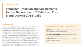 技术公告StemSpan™ Medium and Supplements for the Generation of T Cells from Cord Blood-Derived CD34+ Cells
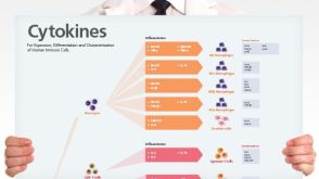 挂图Human Immune Cytokines Infographic of key cytokines for expansion, differentiation and characterization of major immune cell types

 沪公网安备31010102008431号