Vieillard V et al. (AUG 2005)
Proceedings of the National Academy of Sciences 102 31 10981--86
NK cytotoxicity against CD4+ T cells during HIV-1 infection: A gp41 peptide induces the expression of an NKp44 ligand
HIV infection leads to a state of chronic immune activation and progressive deterioration in immune function,manifested most recognizably by the progressive depletion of CD4+ T cells. A substantial percentage of natural killer (NK) cells from patients with HIV infection are activated and express the natural cytotoxicity receptor (NCR) NKp44. Here we show that a cellular ligand for NKp44 (NKp44L) is expressed during HIV-1 infection and is correlated with both the progression of CD4+ T cell depletion and the increase of viral load. CD4+ T cells expressing this ligand are highly sensitive to the NK lysis activity mediated by NKp44+ NK cells. The expression of NKp44L is induced by the linear motif NH2-SWSNKS-COOH of the HIV-1 envelope gp41 protein. This highly conserved motif appears critical to the sharp increase in NK lysis of CD4+ T cells from HIV-infected patients. These studies strongly suggest that induction of NKp44L plays a key role in the lysis of CD4+ T cells by activated NK cells in HIV infection and consequently provide a framework for considering how HIV-1 may use NK cell immune surveillance to trigger CD4+ T cells. Understanding this mechanism may help to develop future therapeutic strategies and vaccines against HIV-1 infection.
View Publication
产品号#:
03800
03801
03802
03803
03804
03805
03806
05150
15021
15061
产品名:
ClonaCell™-HY杂交瘤试剂盒
ClonaCell™-HY培养基A
ClonaCell™-HY 培养基 B
ClonaCell™-HY 培养基 C
ClonaCell™-HY 培养基 D
ClonaCell™-HY 培养基 E
ClonaCell™-HY PEG
MyeloCult™ H5100
RosetteSep™人T细胞富集抗体混合物
RosetteSep™人T细胞富集抗体混合物
Swann J et al. ( 2016)
Virology journal 13 1 30
Cytosolic sulfotransferase 1A1 regulates HIV-1 minus-strand DNA elongation in primary human monocyte-derived macrophages.
BACKGROUND: The cellular sulfonation pathway modulates key steps of virus replication. This pathway comprises two main families of sulfonate-conjugating enzymes: Golgi sulfotransferases,which sulfonate proteins,glycoproteins,glycolipids and proteoglycans; and cytosolic sulfotransferases (SULTs),which sulfonate various small molecules including hormones,neurotransmitters,and xenobiotics. Sulfonation controls the functions of numerous cellular factors such as those involved in cell-cell interactions,cell signaling,and small molecule detoxification. We previously showed that the cellular sulfonation pathway regulates HIV-1 gene expression and reactivation from latency. Here we show that a specific cellular sulfotransferase can regulate HIV-1 replication in primary human monocyte-derived macrophages (MDMs) by yet another mechanism,namely reverse transcription. METHODS: MDMs were derived from monocytes isolated from donor peripheral blood mononuclear cells (PBMCs) obtained from the San Diego Blood Bank. After one week in vitro cell culture under macrophage-polarizing conditions,MDMs were transfected with sulfotranserase-specific or control siRNAs and infected with HIV-1 or SIV constructs expressing a luciferase reporter. Infection levels were subsequently monitored by luminescence. Western blotting was used to assay siRNA knockdown and viral protein levels,and qPCR was used to measure viral RNA and DNA products. RESULTS: We demonstrate that the cytosolic sulfotransferase SULT1A1 is highly expressed in primary human MDMs,and through siRNA knockdown experiments,we show that this enzyme promotes infection of MDMs by single cycle VSV-G pseudotyped human HIV-1 and simian immunodeficiency virus vectors and by replication-competent HIV-1. Quantitative PCR analysis revealed that SULT1A1 affects HIV-1 replication in MDMs by modulating the kinetics of minus-strand DNA elongation during reverse transcription. CONCLUSIONS: These studies have identified SULT1A1 as a cellular regulator of HIV-1 reverse transcription in primary human MDMs. The normal substrates of this enzyme are small phenolic-like molecules,raising the possibility that one or more of these substrates may be involved. Targeting SULT1A1 and/or its substrate(s) may offer a novel host-directed strategy to improve HIV-1 therapeutics.
View Publication
Directed evolution of a recombinase that excises the provirus of most HIV-1 primary isolates with high specificity.
Current combination antiretroviral therapies (cART) efficiently suppress HIV-1 reproduction in humans,but the virus persists as integrated proviral reservoirs in small numbers of cells. To generate an antiviral agent capable of eradicating the provirus from infected cells,we employed 145 cycles of substrate-linked directed evolution to evolve a recombinase (Brec1) that site-specifically recognizes a 34-bp sequence present in the long terminal repeats (LTRs) of the majority of the clinically relevant HIV-1 strains and subtypes. Brec1 efficiently,precisely and safely removes the integrated provirus from infected cells and is efficacious on clinical HIV-1 isolates in vitro and in vivo,including in mice humanized with patient-derived cells. Our data suggest that Brec1 has potential for clinical application as a curative HIV-1 therapy.
View Publication
产品号#:
02697
17896
17896RF
17952
17952RF
21000
20119
20155
04435
04445
100-0696
产品名:
StemSpan™ CC110
EasySep™人脐带血CD34正选试剂盒II
RoboSep™ 人脐带血CD34正选试剂盒II
EasySep™人CD4+ T细胞分选试剂盒
RoboSep™ 人CD4+ T细胞分选试剂盒
RoboSep™- S
RoboSep™ 吸头组件抛光剂
RoboSep™分选管套装(9个塑料管)
MethoCult™ H4435 Enriched
MethoCult™ H4435 Enriched
EasySep™人CD4+ T细胞分离试剂盒
Haase D et al. ( )
Journal of immunotherapy (Hagerstown,Md. : 1997) 38 6 250--8
Large-scale Isolation of Highly Pure Untouched" Regulatory T Cells in a GMP Environment for Adoptive Cell Therapy."
Adoptive cell therapy is an emerging treatment strategy for a number of serious diseases. Regulatory T (Treg) cells represent 1 cell type of particular interest for therapy of inflammatory conditions,as they are responsible for controlling unwanted immune responses. Initial clinical trials of adoptive transfer of Treg cells in patients with graft-versus-host disease were shown to be safe. However,obtaining sufficient numbers of highly pure and functional Treg cells with minimal contamination remains a challenge. We developed a novel approach to isolate untouched" human Treg cells from healthy donors on the basis of negative selection using the surface markers CD49d and CD127. This procedure�
View Publication
产品号#:
18062
18062RF
21000
20119
20155
产品名:
RoboSep™- S
RoboSep™ 吸头组件抛光剂
RoboSep™分选管套装(9个塑料管)
Borsa M et al. ( 2015)
The Virology Journal 12 77
HIV infection and antiretroviral therapy lead to unfolded protein response activation
BACKGROUND: The unfolded protein response (UPR) is one of the pathways triggered to ensure quality control of the proteins assembled in the endoplasmic reticulum (ER) when cell homeostasis is compromised. This mechanism is primarily composed of three transmembrane proteins serving as stress sensors: PKR-like ER kinase (PERK),activating transcription factor 6 (ATF6),and inositol-requiring enzyme 1 (IRE1). These three proteins' synergic action elicits translation and transcriptional downstream pathways,leading to less protein production and activating genes that encode important proteins in folding processes,including chaperones. Previous reports showed that viruses have evolved mechanisms to curtail or customize this UPR signaling for their own benefit. However,HIV infection's effect on the UPR has scarcely been investigated. METHODS: This work investigated UPR modulation by HIV infection by assessing UPR-related protein expression under in vitro and in vivo conditions via Western blotting. Antiretroviral (ARV) drugs' influence on this stress response was also considered. RESULTS: In in vitro and in vivo analyses,our results confirm that HIV infection activates stress-response components and that ARV therapy contributes to changes in the UPR's activation profile. CONCLUSIONS: This is the first report showing UPR-related protein expression in HIV target cells derived directly from HIV-infected patients receiving different ARV therapies. Thus,two mechanisms may occur simultaneously: interference by HIV itself and the ARV drugs' pharmacological effects as UPR activators. New evidence of how HIV modulates the UPR to enhance its own replication and secure infection success is also presented.
View Publication
产品号#:
15022
15062
15028
15068
产品名:
RosetteSep™人CD4+ T细胞富集抗体混合物
RosetteSep™人CD4+ T细胞富集抗体混合物
RosetteSep™人单核细胞富集抗体混合物
RosetteSep™人单核细胞富集抗体混合物
Ohoka Y et al. (JAN 2011)
Journal of immunology (Baltimore,Md. : 1950) 186 2 733--44
Retinoic acid-induced CCR9 expression requires transient TCR stimulation and cooperativity between NFATc2 and the retinoic acid receptor/retinoid X receptor complex.
Retinoic acid (RA) imprints gut-homing specificity on T cells upon activation by inducing the expression of chemokine receptor CCR9 and integrin α4β7. CCR9 expression seemed to be more highly dependent on RA than was the α4β7 expression,but its molecular mechanism remained unclear. In this article,we show that NFAT isoforms NFATc1 and NFATc2 directly interact with RA receptor (RAR) and retinoid X receptor (RXR) but play differential roles in RA-induced CCR9 expression on murine naive CD4(+) T cells. TCR stimulation for 6-24 h was required for the acquisition of responsiveness to RA and induced activation of NFATc1 and NFATc2. However,RA failed to induce CCR9 expression as long as TCR stimulation continued. After terminating TCR stimulation or adding cyclosporin A to the culture,Ccr9 gene transcription was induced,accompanied by inactivation of NFATc1 and sustained activation of NFATc2. Reporter and DNA-affinity precipitation assays demonstrated that the binding of NFATc2 to two NFAT-binding sites and that of the RAR/RXR complex to an RA response element half-site in the 5'-flanking region of the mouse Ccr9 gene were critical for RA-induced promoter activity. NFATc2 directly bound to RARα and RXRα,and it enhanced the binding of RARα to the RA response element half-site. NFATc1 also bound to the NFAT-binding sites and directly to RARα and RXRα,but it inhibited the NFATc2-dependent promoter activity. These results suggest that the cooperativity between NFATc2 and the RAR/RXR complex is essential for CCR9 expression on T cells and that NFATc1 interferes with the action of NFATc2.
View Publication

 EasySep™小鼠TIL(CD45)正选试剂盒


 实验方案Optimizing Delivery Efficiency with Fluorescent Dextran Using the CellPore™ Transfection System
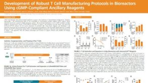 科学海报Development of Robust T Cell Manufacturing Protocols in Bioreactors Using cGMP-Compliant Ancillary Reagents


 沪公网安备31010102008431号