Harlow DE et al. (JAN 2014)
Journal of Neuroscience 34 4 1333--1343
Expression of Proteolipid Protein Gene in Spinal Cord Stem Cells and Early Oligodendrocyte Progenitor Cells Is Dispensable for Normal Cell Migration and Myelination
Plp1 gene expression occurs very early in development,well before the onset of myelination,creating a conundrum with regard to the function of myelin proteolipid protein (PLP),one of the major proteins in compact myelin. Using PLP-EGFP mice to investigate Plp1 promoter activity,we found that,at very early time points,PLP-EGFP was expressed in Sox2+ undifferentiated precursors in the spinal cord ventricular zone (VZ),as well as in the progenitors of both neuronal and glial lineages. As development progressed,most PLP-EGFP-expressing cells gave rise to oligodendrocyte progenitor cells (OPCs). The expression of PLP-EGFP in the spinal cord was quite dynamic during development. PLP-EGFP was highly expressed as cells delaminated from the VZ. Expression was downregulated as cells moved laterally through the cord,and then robustly upregulated as OPCs differentiated into mature myelinating oligodendrocytes. The presence of PLP-EGFP expression in OPCs raises the question of its role in this migratory population. We crossed PLP-EGFP reporter mice into a Plp1-null background to investigate the role of PLP in early OPC development. In the absence of PLP,normal numbers of OPCs were generated and their distribution throughout the spinal cord was unaffected. However,the orientation and length of OPC processes during migration was abnormal in Plp1-null mice,suggesting that PLP plays a role either in the structural integrity of OPC processes or in their response to extracellular cues that orient process outgrowth.
View Publication
产品号#:
05707
产品名:
NeuroCult™化学解离试剂盒(小鼠)
Hou Y et al. (MAY 2014)
Neurobiology of Aging 35 5 975--989
Permeability transition pore-mediated mitochondrial superoxide flashes mediate an early inhibitory effect of amyloid beta1 42 on neural progenitor cell proliferation
Cellular damage by reactive oxygen species and altered neurogenesis are implicated in the etiology of AD and the pathogenic actions of amyloid β-peptide (Aβ); the underlying mechanisms and the early oxidative intracellular events triggered by Aβ are not established. In the present study,we found that mouse embryonic cortical neural progenitor cells exhibit intermittent spontaneous mitochondrial superoxide (SO) flashes that require transient opening of mitochondrial permeability transition pores (mPTPs). The incidence of mitochondria SO flash activity in neural progenitor cells (NPCs) increased during the first 6-24 hours of exposure to aggregating amyloid β-peptide (Aβ1-42),indicating an increase in transient mPTP opening. Subsequently,the SO flash frequency progressively decreased and ceased between 48 and 72 hours of exposure to Aβ1-42,during which time global cellular reactive oxygen species increased,mitochondrial membrane potential decreased,cytochrome C was released from mitochondria and the cells degenerated. Inhibition of mPTPs and selective reduction in mitochondrial SO flashes significantly ameliorated the negative effects of Aβ1-42 on NPC proliferation and survival. Our findings suggest that mPTP-mediated bursts of mitochondrial SO production is a relatively early and pivotal event in the adverse effects of Aβ1-42 on NPCs. If Aβ inhibits NPC proliferation in the brains of AD patients by a similar mechanism,then interventions that inhibit mPTP-mediated superoxide flashes would be expected to protect NPCs against the adverse effects of Aβ.
View Publication
Sox2 expression defines a heterogeneous population of neurosphere-forming cells in the adult murine brain.
The identification of neural stem cells (NSCs) in situ has been prevented by the inability to identify a marker consistently expressed in all adult NSCs and is thus generally accomplished using the in vitro neurosphere-forming assay. The high-mobility group transcription factor Sox2 is expressed in embryonic neural epithelial stem cells; because these cells are thought to give rise to the adult NSC population,we hypothesized that Sox2 may continue to be expressed in adult NSCs. Using Sox2:EGFP transgenic mice,we show that Sox2 is expressed in neurogenic regions along the rostral-caudal axis of the central nervous system throughout life. Furthermore,all neurospheres derived from these neurogenic regions express Sox2,suggesting that Sox2 is indeed expressed in adult NSCs. We demonstrate that NSCs are heterogeneous within the adult brain,with differing capacities for cell production. In vitro,all neurospheres express Sox2,but the expression of markers common to early progenitor cells within individual neurospheres varies; this heterogeneity of NSCs is mirrored in vivo. For example,both glial fibrillary acidic protein and NG2 are expressed within individual neurospheres,but their expression is mutually exclusive; likewise,these two markers show distinct staining patterns within the Sox2+ regions of the brain's neurogenic regions. Thus,we propose that the expression of Sox2 is a unifying characteristic of NSCs in the adult brain,but that not all NSCs maintain the ability to form all neural cell types in vivo.
View Publication
产品号#:
05700
05701
05702
产品名:
NeuroCult™ 基础培养基(小鼠和大鼠)
NeuroCult™ 扩增添加物(小鼠和大鼠)
NeuroCult™扩增试剂盒(小鼠和大鼠)
Fernando P et al. (OCT 2005)
FASEB journal : official publication of the Federation of American Societies for Experimental Biology 19 12 1671--3
Neural stem cell differentiation is dependent upon endogenous caspase 3 activity.
Caspase proteases have become the focal point for the development and application of anti-apoptotic therapies in a variety of central nervous system diseases. However,this approach is based on the premise that caspase function is limited to invoking cell death signals. Here,we show that caspase-3 activity is elevated in nonapoptotic differentiating neuronal cell populations. Moreover,peptide inhibition of protease activity effectively inhibits the differentiation process in a cultured neurosphere model. These results implicate caspase-3 activation as a conserved feature of neuronal differentiation and suggest that targeted inhibition of this protease in neural cell populations may have unintended consequences.
View Publication
产品号#:
05700
05701
05702
05703
05704
产品名:
NeuroCult™ 基础培养基(小鼠和大鼠)
NeuroCult™ 扩增添加物(小鼠和大鼠)
NeuroCult™扩增试剂盒(小鼠和大鼠)
NeuroCult™ 分化添加物(小鼠和大鼠)
NeuroCult™ 分化试剂盒(小鼠和大鼠)
Young KM et al. (AUG 2007)
The Journal of neuroscience : the official journal of the Society for Neuroscience 27 31 8286--96
Subventricular zone stem cells are heterogeneous with respect to their embryonic origins and neurogenic fates in the adult olfactory bulb.
We determined the embryonic origins of adult forebrain subventricular zone (SVZ) stem cells by Cre-lox fate mapping in transgenic mice. We found that all parts of the telencephalic neuroepithelium,including the medial ganglionic eminence and lateral ganglionic eminence (LGE) and the cerebral cortex,contribute multipotent,self-renewing stem cells to the adult SVZ. Descendants of the embryonic LGE and cortex settle in ventral and dorsal aspects of the dorsolateral SVZ,respectively. Both populations contribute new (5-bromo-2'-deoxyuridine-labeled) tyrosine hydroxylase- and calretinin-positive interneurons to the adult olfactory bulb. However,calbindin-positive interneurons in the olfactory glomeruli were generated exclusively by LGE-derived stem cells. Thus,different SVZ stem cells have different embryonic origins,colonize different parts of the SVZ,and generate different neuronal progeny,suggesting that some aspects of embryonic patterning are preserved in the adult SVZ. This could have important implications for the design of endogenous stem cell-based therapies in the future.
View Publication
Aberrant Neural Stem Cell Proliferation and Increased Adult Neurogenesis in Mice Lacking Chromatin Protein HMGB2
Neural stem and progenitor cells (NSCs/NPCs) are distinct groups of cells found in the mammalian central nervous system (CNS). Previously we determined that members of the High Mobility Group (HMG) B family of chromatin structural proteins modulate NSC proliferation and self-renewal. Among them HMGB2 was found to be dynamically expressed in proliferating and differentiating NSCs,suggesting that it may regulate NSC maintenance. We report now that Hmgb2(-/-) mice exhibit SVZ hyperproliferation,increased numbers of SVZ NSCs,and a trend towards aberrant increases in newly born neurons in the olfactory bulb (OB) granule cell layer. Increases in the levels of the transcription factor p21 and the Neural cell adhesion molecule (NCAM),along with down-regulation of the transcription/pluripotency factor Oct4 in the Hmgb2-/- SVZ point to a possible pathway for this increased proliferation/differentiation. Our findings suggest that HMGB2 functions as a modulator of neurogenesis in young adult mice through regulation of NSC proliferation,and identify a potential target via which CNS repair could be amplified following trauma or disease-based neuronal degeneration.
View Publication
产品号#:
05700
05701
05702
05715
产品名:
NeuroCult™ 基础培养基(小鼠和大鼠)
NeuroCult™ 扩增添加物(小鼠和大鼠)
NeuroCult™扩增试剂盒(小鼠和大鼠)
NeuroCult™成年中枢神经系统(CNS)组织酶解试剂盒(小鼠和大鼠)
Ji M et al. (SEP 2013)
Science Translational Medicine 5 201 201ra119--201ra119
Rapid, Label-Free Detection of Brain Tumors with Stimulated Raman Scattering Microscopy
Surgery is an essential component in the treatment of brain tumors. However,delineating tumor from normal brain remains a major challenge. We describe the use of stimulated Raman scattering (SRS) microscopy for differentiating healthy human and mouse brain tissue from tumor-infiltrated brain based on histoarchitectural and biochemical differences. Unlike traditional histopathology,SRS is a label-free technique that can be rapidly performed in situ. SRS microscopy was able to differentiate tumor from nonneoplastic tissue in an infiltrative human glioblastoma xenograft mouse model based on their different Raman spectra. We further demonstrated a correlation between SRS and hematoxylin and eosin microscopy for detection of glioma infiltration (κ = 0.98). Finally,we applied SRS microscopy in vivo in mice during surgery to reveal tumor margins that were undetectable under standard operative conditions. By providing rapid intraoperative assessment of brain tissue,SRS microscopy may ultimately improve the safety and accuracy of surgeries where tumor boundaries are visually indistinct.
View Publication


 EasySep™小鼠TIL(CD45)正选试剂盒




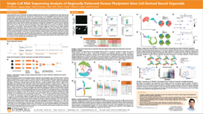 科学海报Single-Cell RNA Sequencing Analysis of Regionally Patterned Human Pluripotent Stem Cell-Derived Neural Organoids
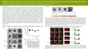 科学海报STEMdiff™ Cerebral Organoid Kit: A New Tool for the Culture of 3D Brain Organoids Derived from hPSCs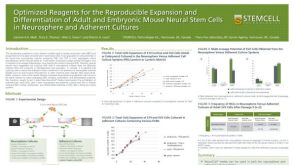


 沪公网安备31010102008431号