Ito N et al. (APR 2016)
Disease models & mechanisms 9 4 451--462
Decreased N-TAF1 expression in X-linked dystonia-parkinsonism patient-specific neural stem cells.
X-linked dystonia-parkinsonism (XDP) is a hereditary neurodegenerative disorder involving a progressive loss of striatal medium spiny neurons. The mechanisms underlying neurodegeneration are not known,in part because there have been few cellular models available for studying the disease. The XDP haplotype consists of multiple sequence variations in a region of the X chromosome containingTAF1,a large gene with at least 38 exons,and a multiple transcript system (MTS) composed of five unconventional exons. A previous study identified an XDP-specific insertion of a SINE-VNTR-Alu (SVA)-type retrotransposon in intron 32 ofTAF1,as well as a neural-specific TAF1 isoform,N-TAF1,which showed decreased expression in post-mortem XDP brain compared with control tissue. Here,we generated XDP patient and control fibroblasts and induced pluripotent stem cells (iPSCs) in order to further probe cellular defects associated with this disease. As initial validation of the model,we compared expression ofTAF1and MTS transcripts in XDP versus control fibroblasts and iPSC-derived neural stem cells (NSCs). Compared with control cells,XDP fibroblasts exhibited decreased expression ofTAF1transcript fragments derived from exons 32-36,a region spanning the SVA insertion site. N-TAF1,which incorporates an alternative exon (exon 34'),was not expressed in fibroblasts,but was detectable in iPSC-differentiated NSCs at levels that were ∼threefold lower in XDP cells than in controls. These results support the previous findings that N-TAF1 expression is impaired in XDP,but additionally indicate that this aberrant transcription might occur in neural cells at relatively early stages of development that precede neurodegeneration.
View Publication
产品号#:
产品名:
Sancho-Martinez I et al. (FEB 2016)
Nature communications 7 10743
Establishment of human iPSC-based models for the study and targeting of glioma initiating cells.
Glioma tumour-initiating cells (GTICs) can originate upon the transformation of neural progenitor cells (NPCs). Studies on GTICs have focused on primary tumours from which GTICs could be isolated and the use of human embryonic material. Recently,the somatic genomic landscape of human gliomas has been reported. RTK (receptor tyrosine kinase) and p53 signalling were found dysregulated in ∼90% and 86% of all primary tumours analysed,respectively. Here we report on the use of human-induced pluripotent stem cells (hiPSCs) for modelling gliomagenesis. Dysregulation of RTK and p53 signalling in hiPSC-derived NPCs (iNPCs) recapitulates GTIC properties in vitro. In vivo transplantation of transformed iNPCs leads to highly aggressive tumours containing undifferentiated stem cells and their differentiated derivatives. Metabolic modulation compromises GTIC viability. Last,screening of 101 anti-cancer compounds identifies three molecules specifically targeting transformed iNPCs and primary GTICs. Together,our results highlight the potential of hiPSCs for studying human tumourigenesis.
View Publication
产品号#:
05850
05857
05870
05875
85850
85857
85870
85875
产品名:
mTeSR™1
mTeSR™1
Ostrakhovitch EA et al. (DEC 2012)
Archives of biochemistry and biophysics 528 1 21--31
Directed differentiation of embryonic P19 cells and neural stem cells into neural lineage on conducting PEDOT-PEG and ITO glass substrates.
Differentiation of pluripotent and lineage restricted stem cells such as neural stem cells (NSCs) was studied on conducting substrates of various nature without perturbation of the genome with exogenous genetic material or chemical stimuli. Primary mouse adult neural stem cells (NSCs) and P19 pluripotent embryonal (P19 EC) carcinoma cells were used. Expression levels of neuronal markers β-III-tubulin and neurofilament were evaluated by immunochemistry and flow cytometry. It was shown that the ability of the substrate to induce differentiation directly correlated with its conductivity. Conducting substrates (conducting oxides or doped pi-conjugated organic polymers) with different morphology,structure,and conductivity mechanisms all promoted differentiation of NSC and P19 cells into neuronal lineage to a similar degree without use of additional factors such as poly-L-ornithine coating or retinoic acid,as verified by their morphology and upregulation of the neuronal markers but not astrocyte marker GFAP. However,substrates with low conductance below ca. 10(-4) S cm(-2) did not show this ability. Morphology of differentiating cells was visualized by atomic force microscopy. NSCs cells increased β-III-tubulin expression by 95% and P19 cells by over 30%. Our results suggest that the substrate conductivity is a key factor governing the cell fate. Differentiation of P19 cells into neuronal lineage on conducting substrates was attributed to downregualtion of Akt signaling pathway and increase in expression of dual oxidase 1 (DUOX 1).
View Publication
产品号#:
05700
05701
05702
05703
05704
05715
产品名:
NeuroCult™ 基础培养基(小鼠和大鼠)
NeuroCult™ 扩增添加物(小鼠和大鼠)
NeuroCult™扩增试剂盒(小鼠和大鼠)
NeuroCult™ 分化添加物(小鼠和大鼠)
NeuroCult™ 分化试剂盒(小鼠和大鼠)
NeuroCult™成年中枢神经系统(CNS)组织酶解试剂盒(小鼠和大鼠)
Belkind-Gerson J et al. (JAN 2013)
Neurogastroenterology and motility : the official journal of the European Gastrointestinal Motility Society 25 1 61--9.e7
Nestin-expressing cells in the gut give rise to enteric neurons and glial cells.
BACKGROUND Neuronal stem cells (NSCs) are promising for neurointestinal disease therapy. Although NSCs have been isolated from intestinal musclularis,their presence in mucosa has not been well described. Mucosa-derived NSCs are accessible endoscopically and could be used autologously. Brain-derived Nestin-positive NSCs are important in endogenous repair and plasticity. The aim was to isolate and characterize mucosa-derived NSCs,determine their relationship to Nestin-expressing cells and to demonstrate their capacity to produce neuroglial networks in vitro and in vivo. METHODS Neurospheres were generated from periventricular brain,colonic muscularis (Musc),and mucosa-submucosa (MSM) of mice expressing green fluorescent protein (GFP) controlled by the Nestin promoter (Nestin-GFP). Neuronal stem cells were also grown as adherent colonies from intestinal mucosal organoids. Their differentiation potential was assessed using immunohistochemistry using glial and neuronal markers. Brain and gut-derived neurospheres were transplanted into explants of chick embryonic aneural hindgut to determine their fate. KEY RESULTS Musc- and MSM-derived neurospheres expressed Nestin and gave rise to cells of neuronal,glial,and mesenchymal lineage. Although Nestin expression in tissue was mostly limited to glia co-labelled with glial fibrillary acid protein (GFAP),neurosphere-derived neurons and glia both expressed Nestin in vitro,suggesting that Nestin+/GFAP+ glial cells may give rise to new neurons. Moreover,following transplantation into aneural colon,brain- and gut-derived NSCs were able to differentiate into neurons. CONCLUSIONS & INFERENCES Nestin-expressing intestinal NSCs cells give rise to neurospheres,differentiate into neuronal,glial,and mesenchymal lineages in vitro,generate neurons in vivo and can be isolated from mucosa. Further studies are needed for exploring their potential for treating neuropathies.
View Publication
产品号#:
05700
05701
05702
05703
05704
05715
产品名:
NeuroCult™ 基础培养基(小鼠和大鼠)
NeuroCult™ 扩增添加物(小鼠和大鼠)
NeuroCult™扩增试剂盒(小鼠和大鼠)
NeuroCult™ 分化添加物(小鼠和大鼠)
NeuroCult™ 分化试剂盒(小鼠和大鼠)
NeuroCult™成年中枢神经系统(CNS)组织酶解试剂盒(小鼠和大鼠)
Maynard KR and Stein E (NOV 2012)
The Journal of neuroscience : the official journal of the Society for Neuroscience 32 47 16637--50
DSCAM contributes to dendrite arborization and spine formation in the developing cerebral cortex.
Down syndrome cell adhesion molecule,or DSCAM,has been implicated in many neurodevelopmental processes including axon guidance,dendrite arborization,and synapse formation. Here we show that DSCAM plays an important role in regulating the morphogenesis of cortical pyramidal neurons in the mouse. We report that DSCAM expression is developmentally regulated and localizes to synaptic plasma membranes during a time of robust cortical dendrite arborization and spine formation. Analysis of mice that carry a spontaneous mutation in DSCAM (DSCAM(del17)) revealed gross morphological changes in brain size and shape in addition to subtle changes in cortical organization,volume,and lamination. Early postnatal mutant mice displayed a transient decrease in cortical thickness,but these reductions could not be attributed to changes in neuron production or cell death. DSCAM(del17) mutants showed temporary impairments in the branching of layer V pyramidal neuron dendrites at P10 and P17 that recovered to normal by adulthood. Defects in DSCAM(del17) dendrite branching correlated with a temporal increase in apical branch spine density and lasting changes in spine morphology. At P15 and P42,mutant mice displayed a decrease in the percentage of large,stable spines and an increase in the percentage of small,immature spines. Together,our findings suggest that DSCAM contributes to pyramidal neuron morphogenesis by regulating dendrite arborization and spine formation during cortical circuit development.
View Publication
产品号#:
05715
产品名:
NeuroCult™成年中枢神经系统(CNS)组织酶解试剂盒(小鼠和大鼠)
Conte D et al. (JAN 2012)
PloS one 7 12 e52167
Loss of Atrx sensitizes cells to DNA damaging agents through p53-mediated death pathways.
Prevalent cell death in forebrain- and Sertoli cell-specific Atrx knockout mice suggest that Atrx is important for cell survival. However,conditional ablation in other tissues is not associated with increased death indicating that diverse cell types respond differently to the loss of this chromatin remodeling protein. Here,primary macrophages isolated from Atrx(f/f) mice were infected with adenovirus expressing Cre recombinase or β-galactosidase,and assayed for cell survival under different experimental conditions. Macrophages survive without Atrx but undergo rapid apoptosis upon lipopolysaccharide (LPS) activation suggesting that chromatin reorganization in response to external stimuli is compromised. Using this system we next tested the effect of different apoptotic stimuli on cell survival. We observed that survival of Atrx-null cells were similar to wild type cells in response to serum withdrawal,anti-Fas antibody,C2 ceramide or dexamethasone treatment but were more sensitive to 5-fluorouracil (5-FU). Cell survival could be rescued by re-introducing Atrx or by removal of p53 demonstrating the cell autonomous nature of the effect and its p53-dependence. Finally,we demonstrate that multiple primary cell types (myoblasts,embryonic fibroblasts and neurospheres) were sensitive to 5-FU,cisplatin,and UV light treatment. Together,our results suggest that cells lacking Atrx are more sensitive to DNA damaging agents and that this may result in enhanced death during development when cells are at their proliferative peak. Moreover,it identifies potential treatment options for cancers associated with ATRX mutations,including glioblastoma and pancreatic neuroendocrine tumors.
View Publication
Handel AE et al. (MAR 2016)
Human Molecular Genetics 25 5 989--1000
Assessing similarity to primary tissue and cortical layer identity in induced pluripotent stem cell-derived cortical neurons through single-cell transcriptomics
Induced pluripotent stem cell (iPSC)-derived cortical neurons potentially present a powerful new model to understand corticogenesis and neurological disease. Previous work has established that differentiation protocols can produce cortical neurons,but little has been done to characterize these at cellular resolution. In particular,it is unclear to what extent in vitro two-dimensional,relatively disordered culture conditions recapitulate the development of in vivo cortical layer identity. Single-cell multiplex reverse transcriptase-quantitative polymerase chain reaction (RT-qPCR) was used to interrogate the expression of genes previously implicated in cortical layer or phenotypic identity in individual cells. Totally,93.6% of single cells derived from iPSCs expressed genes indicative of neuronal identity. High proportions of single neurons derived from iPSCs expressed glutamatergic receptors and synaptic genes. And,68.4% of iPSC-derived neurons expressing at least one layer marker could be assigned to a laminar identity using canonical cortical layer marker genes. We compared single-cell RNA-seq of our iPSC-derived neurons to available single-cell RNA-seq data from human fetal and adult brain and found that iPSC-derived cortical neurons closely resembled primary fetal brain cells. Unexpectedly,a subpopulation of iPSC-derived neurons co-expressed canonical fetal deep and upper cortical layer markers. However,this appeared to be concordant with data from primary cells. Our results therefore provide reassurance that iPSC-derived cortical neurons are highly similar to primary cortical neurons at the level of single cells but suggest that current layer markers,although effective,may not be able to disambiguate cortical layer identity in all cells.
View Publication

 EasySep™小鼠TIL(CD45)正选试剂盒


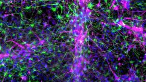 实验方案How to Co-Culture Astrocytes and NGN2 mRNA-Driven Induced Forebrain Neurons Derived from Human Pluripotent Stem Cells

 沪公网安备31010102008431号