Bhinge A et al. (JUN 2014)
EMBO Journal 33 11 1271--1283
MiR-135b is a direct PAX6 target and specifies human neuroectoderm by inhibiting TGF-$\$/BMP signaling.
Several transcription factors (TFs) have been implicated in neuroectoderm (NE) development,and recently,the TF PAX6 was shown to be critical for human NE specification. However,microRNA networks regulating human NE development have been poorly documented. We hypothesized that microRNAs activated by PAX6 should promote NE development. Using a genomics approach,we identified PAX6 binding sites and active enhancers genome-wide in an in vitro model of human NE development that was based on neural differentiation of human embryonic stem cells (hESC). PAX6 binding to active enhancers was found in the proximity of several microRNAs,including hsa-miR-135b. MiR-135b was activated during NE development,and ectopic expression of miR-135b in hESC promoted differentiation toward NE. MiR-135b promotes neural conversion by targeting components of the TGF-β and BMP signaling pathways,thereby inhibiting differentiation into alternate developmental lineages. Our results demonstrate a novel TF-miRNA module that is activated during human neuroectoderm development and promotes the irreversible fate specification of human pluripotent cells toward the neural lineage.
View Publication
产品号#:
05850
05857
05870
05875
85850
85857
85870
85875
产品名:
mTeSR™1
mTeSR™1
Lippmann ES et al. (FEB 2014)
Scientific reports 4 February 2014 4160
A retinoic acid-enhanced, multicellular human blood-brain barrier model derived from stem cell sources.
Blood-brain barrier (BBB) models are often used to investigate BBB function and screen brain-penetrating therapeutics,but it has been difficult to construct a human model that possesses an optimal BBB phenotype and is readily scalable. To address this challenge,we developed a human in vitro BBB model comprising brain microvascular endothelial cells (BMECs),pericytes,astrocytes and neurons derived from renewable cell sources. First,retinoic acid (RA) was used to substantially enhance BBB phenotypes in human pluripotent stem cell (hPSC)-derived BMECs,particularly through adherens junction,tight junction,and multidrug resistance protein regulation. RA-treated hPSC-derived BMECs were subsequently co-cultured with primary human brain pericytes and human astrocytes and neurons derived from human neural progenitor cells (NPCs) to yield a fully human BBB model that possessed significant tightness as measured by transendothelial electrical resistance (˜5,000 $\$(2)). Overall,this scalable human BBB model may enable a wide range of neuroscience studies.
View Publication
产品号#:
05850
05857
05870
05875
85850
85857
85870
85875
产品名:
mTeSR™1
mTeSR™1
Lippmann ES et al. (APR 2014)
Stem Cells 32 4 1032--1042
Defined human pluripotent stem cell culture enables highly efficient neuroepithelium derivation without small molecule inhibitors.
The embryonic neuroepithelium gives rise to the entire central nervous system in vivo,making it an important tissue for developmental studies and a prospective cell source for regenerative applications. Current protocols for deriving homogenous neuroepithelial cultures from human pluripotent stem cells (hPSCs) consist of either embryoid body-mediated neuralization followed by a manual isolation step or adherent differentiation using small molecule inhibitors. Here,we report that hPSCs maintained under chemically defined,feeder-independent,and xeno-free conditions can be directly differentiated into pure neuroepithelial cultures ([mt]90% Pax6(+)/N-cadherin(+) with widespread rosette formation) within 6 days under adherent conditions,without small molecule inhibitors,and using only minimalistic medium consisting of Dulbecco's modified Eagle's medium/F-12,sodium bicarbonate,selenium,ascorbic acid,transferrin,and insulin (i.e.,E6 medium). Furthermore,we provide evidence that the defined culture conditions enable this high level of neural conversion in contrast to hPSCs maintained on mouse embryonic fibroblasts (MEFs). In addition,hPSCs previously maintained on MEFs could be rapidly converted to a neural compliant state upon transfer to these defined conditions while still maintaining their ability to generate all three germ layers. Overall,this fully defined and scalable protocol should be broadly useful for generating therapeutic neural cells for regenerative applications.
View Publication
Overexpression of calcium-permeable glutamate receptors in glioblastoma derived brain tumor initiating cells.
Glioblastoma multiforme is the most malignant type of primary brain tumor with a poor prognosis. These tumors consist of a heterogeneous population of malignant cells,including well-differentiated tumor cells and less differentiated cells with stem cell properties. These cancer stem cells,known as brain tumor initiating cells,likely contribute to glioma recurrence,as they are highly invasive,mobile,resistant to radiation and chemotherapy,and have the capacity to self-renew. Glioblastoma tumor cells release excitotoxic levels of glutamate,which may be a key process in the death of peritumoral neurons,formation of necrosis,local inflammation,and glioma-related seizures. Moreover,elevated glutamate levels in the tumor may act in paracrine and autocrine manner to activate glutamate receptors on glioblastoma tumor cells,resulting in proliferation and invasion. Using a previously described culturing condition that selectively promotes the growth of brain tumor initiating cells,which express the stem cell markers nestin and SOX-2,we characterize the expression of α-amino-3-hydroxy-5-methyl-4-isozolepropionic acid (AMPA)-type glutamate receptor subunits in brain tumor initiating cells derived from glioblastomas. Here we show for the first time that glioblastoma brain tumor initiating cells express high concentrations of functional calcium-permeable AMPA receptors,compared to the differentiated tumor cultures consisting of non-stem cells. Up-regulated calcium-permeable AMPA receptor expression was confirmed by immunoblotting,immunocytochemistry,and intracellular calcium imaging in response to specific agonists. Our findings raise the possibility that glutamate secretion in the GBM tumor microenvironment may stimulate brain tumor derived cancer stem cells.
View Publication
产品号#:
05750
产品名:
NeuroCult™ NS-A 基础培养基(人)
Saharan S et al. (MAY 2013)
Journal of Neuroscience Research 91 5 642--659
SIRT1 regulates the neurogenic potential of neural precursors in the adult subventricular zone and hippocampus
Within the two neurogenic niches of the adult mammalian brain,i.e.,the subventricular zone lining the lateral ventricle and the subgranular zone of the hippocampus,there exist distinct populations of proliferating neural precursor cells that differentiate to generate new neurons. Numerous studies have suggested that epigenetic regulation by histone-modifying proteins is important in guiding precursor differentiation during development; however,the role of these proteins in regulating neural precursor activity in the adult neurogenic niches remains poorly understood. Here we examine the role of an NAD(+) -dependent histone deacetylase,SIRT1,in modulating the neurogenic potential of neural precursors in the neurogenic niches of the adult mouse brain. We show that SIRT1 is expressed by proliferating adult subventricular zone and hippocampal neural precursors,although its transcript and protein levels are dramatically reduced during neural precursor differentiation. Utilizing a lentiviral-mediated delivery strategy,we demonstrate that abrogation of SIRT1 signaling by RNAi does not affect neural precursor numbers or their proliferation. However,SIRT1 knock down results in a significant increase in neuronal production in both the subventricular zone and the hippocampus. In contrast,enhancing SIRT1 signaling either through lentiviral-mediated SIRT1 overexpression or through use of the SIRT1 chemical activator Resveratrol prevents adult neural precursors from differentiating into neurons. Importantly,knock down of SIRT1 in hippocampal precursors in vivo,either through RNAi or through genetic ablation,promotes their neurogenic potential. These findings highlight SIRT1 signaling as a negative regulator of neuronal differentiation of adult subventricular zone and hippocampal neural precursors. textcopyright 2013 Wiley Periodicals,Inc.
View Publication
产品号#:
05700
05701
05702
产品名:
NeuroCult™ 基础培养基(小鼠和大鼠)
NeuroCult™ 扩增添加物(小鼠和大鼠)
NeuroCult™扩增试剂盒(小鼠和大鼠)
Belkind-Gerson J et al. (JAN 2013)
Neurogastroenterology and motility : the official journal of the European Gastrointestinal Motility Society 25 1 61--9.e7
Nestin-expressing cells in the gut give rise to enteric neurons and glial cells.
BACKGROUND Neuronal stem cells (NSCs) are promising for neurointestinal disease therapy. Although NSCs have been isolated from intestinal musclularis,their presence in mucosa has not been well described. Mucosa-derived NSCs are accessible endoscopically and could be used autologously. Brain-derived Nestin-positive NSCs are important in endogenous repair and plasticity. The aim was to isolate and characterize mucosa-derived NSCs,determine their relationship to Nestin-expressing cells and to demonstrate their capacity to produce neuroglial networks in vitro and in vivo. METHODS Neurospheres were generated from periventricular brain,colonic muscularis (Musc),and mucosa-submucosa (MSM) of mice expressing green fluorescent protein (GFP) controlled by the Nestin promoter (Nestin-GFP). Neuronal stem cells were also grown as adherent colonies from intestinal mucosal organoids. Their differentiation potential was assessed using immunohistochemistry using glial and neuronal markers. Brain and gut-derived neurospheres were transplanted into explants of chick embryonic aneural hindgut to determine their fate. KEY RESULTS Musc- and MSM-derived neurospheres expressed Nestin and gave rise to cells of neuronal,glial,and mesenchymal lineage. Although Nestin expression in tissue was mostly limited to glia co-labelled with glial fibrillary acid protein (GFAP),neurosphere-derived neurons and glia both expressed Nestin in vitro,suggesting that Nestin+/GFAP+ glial cells may give rise to new neurons. Moreover,following transplantation into aneural colon,brain- and gut-derived NSCs were able to differentiate into neurons. CONCLUSIONS & INFERENCES Nestin-expressing intestinal NSCs cells give rise to neurospheres,differentiate into neuronal,glial,and mesenchymal lineages in vitro,generate neurons in vivo and can be isolated from mucosa. Further studies are needed for exploring their potential for treating neuropathies.
View Publication
产品号#:
05700
05701
05702
05703
05704
05715
产品名:
NeuroCult™ 基础培养基(小鼠和大鼠)
NeuroCult™ 扩增添加物(小鼠和大鼠)
NeuroCult™扩增试剂盒(小鼠和大鼠)
NeuroCult™ 分化添加物(小鼠和大鼠)
NeuroCult™ 分化试剂盒(小鼠和大鼠)
NeuroCult™成年中枢神经系统(CNS)组织酶解试剂盒(小鼠和大鼠)
Maynard KR and Stein E (NOV 2012)
The Journal of neuroscience : the official journal of the Society for Neuroscience 32 47 16637--50
DSCAM contributes to dendrite arborization and spine formation in the developing cerebral cortex.
Down syndrome cell adhesion molecule,or DSCAM,has been implicated in many neurodevelopmental processes including axon guidance,dendrite arborization,and synapse formation. Here we show that DSCAM plays an important role in regulating the morphogenesis of cortical pyramidal neurons in the mouse. We report that DSCAM expression is developmentally regulated and localizes to synaptic plasma membranes during a time of robust cortical dendrite arborization and spine formation. Analysis of mice that carry a spontaneous mutation in DSCAM (DSCAM(del17)) revealed gross morphological changes in brain size and shape in addition to subtle changes in cortical organization,volume,and lamination. Early postnatal mutant mice displayed a transient decrease in cortical thickness,but these reductions could not be attributed to changes in neuron production or cell death. DSCAM(del17) mutants showed temporary impairments in the branching of layer V pyramidal neuron dendrites at P10 and P17 that recovered to normal by adulthood. Defects in DSCAM(del17) dendrite branching correlated with a temporal increase in apical branch spine density and lasting changes in spine morphology. At P15 and P42,mutant mice displayed a decrease in the percentage of large,stable spines and an increase in the percentage of small,immature spines. Together,our findings suggest that DSCAM contributes to pyramidal neuron morphogenesis by regulating dendrite arborization and spine formation during cortical circuit development.
View Publication

 EasySep™小鼠TIL(CD45)正选试剂盒


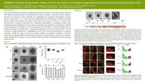 科学海报STEMdiff™ Cerebral Organoid Kit: A New Tool for the Culture of 3D Brain Organoids Derived from hPSCs


 沪公网安备31010102008431号