Abuljadayel IS (JAN 2003)
Current medical research and opinion 19 5 355--75
Induction of stem cell-like plasticity in mononuclear cells derived from unmobilised adult human peripheral blood.
Undifferentiated pluripotent stem cells with flexible developmental potentials are not normally found in peripheral blood. However,such cells have recently been reported to reside in the bone marrow. Herein are reported methods of inducing pluripotency in cells derived from unmobilised adult human peripheral blood. In response to the inclusion of purified CR3/43 monoclonal antibody (mAb) to well-established culture conditions,mononuclear cells (MNC) obtained from a single blood donor are converted into pluripotent haematopoietic,neuronal and cardiomyogenic progenitor stem cells or undifferentiated stem cells. The haematopoietic stem cells are CD34+,clonogenic and have been shown to repopulate non-obese diabetic/severe combined immunodeficient (NOD/SCID) mice. The neuronal precursors transcribe the primitive stem cell markers OCT-4 and nestin,and on maturation,differentially stain positive for neuronal,glial or oligodendrocyte-specific antigens. The cardiomyogenic progenitor stem cells form large bodies of asynchronously beating cells and differentiate into mature cardiomyocytes which transcribe GATA-4. The undifferentiated stem cells do not express haematopoietic-associated markers,are negative for major histocompatibility complex (MHC) class I and II antigens,transcribe high levels of OCT-4 and form embryoid body (EB)-like structures. This induction of stem cell-like plasticity in MNC may have proceeded by a process of retrodifferentiation but,in any case,could have profound clinical and pharmacological implications. Finally,the flexibility and the speed by which a variety of stem cell classes can be generated ex vivo from donor blood could potentially transfer this novel process into a less invasive automated clinical procedure.
View Publication
产品号#:
04434
04444
产品名:
MethoCult™ H4434 Classic
MethoCult™ H4434 Classic
Cheng L et al. (JUN 2014)
Cell Research 24 6 665--679
Generation of neural progenitor cells by chemical cocktails and hypoxia
Neural progenitor cells (NPCs) can be induced from somatic cells by defined factors. Here we report that NPCs can be generated from mouse embryonic fibroblasts by a chemical cocktail,namely VCR (V,VPA,an inhibitor of HDACs; C,CHIR99021,an inhibitor of GSK-3 kinases and R,Repsox,an inhibitor of TGF-β pathways),under a physiological hypoxic condition. These chemical-induced NPCs (ciNPCs) resemble mouse brain-derived NPCs re- garding their proliferative and self-renewing abilities,gene expression profiles,and multipotency for different neu- roectodermal lineages in vitro and in vivo. Further experiments reveal that alternative cocktails with inhibitors of histone deacetylation,glycogen synthase kinase,and TGF-β pathways show similar efficacies for ciNPC induction. Moreover,ciNPCs can also be induced from mouse tail-tip fibroblasts and human urinary cells with the same chemi- cal cocktail VCR. Thus our study demonstrates that lineage-specific conversion of somatic cells to NPCs could be achieved by chemical cocktails without introducing exogenous factors.
View Publication
产品号#:
05850
05857
05870
05875
85850
85857
85870
85875
产品名:
mTeSR™1
mTeSR™1
Kishigami S et al. (FEB 2006)
Biochemical and biophysical research communications 340 1 183--9
Significant improvement of mouse cloning technique by treatment with trichostatin A after somatic nuclear transfer.
The low success rate of animal cloning by somatic cell nuclear transfer (SCNT) is believed to be associated with epigenetic errors including abnormal DNA hypermethylation. Recently,we elucidated by using round spermatids that,after nuclear transfer,treatment of zygotes with trichostatin A (TSA),an inhibitor of histone deacetylase,can remarkably reduce abnormal DNA hypermethylation depending on the origins of transferred nuclei and their genomic regions [S. Kishigami,N. Van Thuan,T. Hikichi,H. Ohta,S. Wakayama. E. Mizutani,T. Wakayama,Epigenetic abnormalities of the mouse paternal zygotic genome associated with microinsemination of round spermatids,Dev. Biol. (2005) in press]. Here,we found that 5-50 nM TSA-treatment for 10 h following oocyte activation resulted in more efficient in vitro development of somatic cloned embryos to the blastocyst stage from 2- to 5-fold depending on the donor cells including tail tip cells,spleen cells,neural stem cells,and cumulus cells. This TSA-treatment also led to more than 5-fold increase in success rate of mouse cloning from cumulus cells without obvious abnormality but failed to improve ES cloning success. Further,we succeeded in establishment of nuclear transfer-embryonic stem (NT-ES) cells from TSA-treated cloned blastocyst at a rate three times higher than those from untreated cloned blastocysts. Thus,our data indicate that TSA-treatment after SCNT in mice can dramatically improve the practical application of current cloning techniques.
View Publication
产品号#:
05700
05701
05702
72282
72284
产品名:
NeuroCult™ 基础培养基(小鼠和大鼠)
NeuroCult™ 扩增添加物(小鼠和大鼠)
NeuroCult™扩增试剂盒(小鼠和大鼠)
曲古抑菌素 A(Trichostatin A)
曲古抑菌素 A(Trichostatin A)
Zhang Z et al. (JAN 2006)
Human molecular genetics 15 2 337--46
Palmitoyl-protein thioesterase-1 deficiency mediates the activation of the unfolded protein response and neuronal apoptosis in INCL.
Numerous proteins undergo modification by palmitic acid (S-acylation) for their biological functions including signal transduction,vesicular transport and maintenance of cellular architecture. Although palmitoylation is an essential modification,these proteins must also undergo depalmitoylation for their degradation by lysosomal proteases. Palmitoyl-protein thioesterase-1 (PPT1),a lysosomal enzyme,cleaves thioester linkages in S-acylated proteins and removes palmitate residues facilitating the degradation of these proteins. Thus,inactivating mutations in the PPT1 gene cause infantile neuronal ceroid lipofuscinosis (INCL),a devastating neurodegenerative storage disorder of childhood. Although rapidly progressing brain atrophy is the most dramatic pathological manifestation of INCL,the molecular mechanism(s) remains unclear. Using PPT1-knockout (PPT1-KO) mice that mimic human INCL,we report here that the endoplasmic reticulum (ER) in the brain cells of these mice is structurally abnormal. Further,we demonstrate that the level of growth-associated protein-43 (GAP-43),a palmitoylated neuronal protein,is elevated in the brains of PPT1-KO mice. Moreover,forced expression of GAP-43 in PPT1-deficient cells results in the abnormal accumulation of this protein in the ER. Consistent with these results,we found evidence for the activation of unfolded protein response (UPR) marked by elevated levels of phosphorylated translation initiation factor,eIF2alpha,increased expression of chaperone proteins such as glucose-regulated protein-78 and activation of caspase-12,a cysteine proteinase in the ER,mediating caspase-3 activation and apoptosis. Our results,for the first time,link PPT1 deficiency with the activation of UPR,apoptosis and neurodegeneration in INCL and identify potential targets for therapeutic intervention in this uniformly fatal disease.
View Publication
产品号#:
05700
05701
05702
产品名:
NeuroCult™ 基础培养基(小鼠和大鼠)
NeuroCult™ 扩增添加物(小鼠和大鼠)
NeuroCult™扩增试剂盒(小鼠和大鼠)
Li J-M et al. (FEB 2007)
Molecular endocrinology (Baltimore,Md.) 21 2 499--511
Angiotensin II-induced neural differentiation via angiotensin II type 2 (AT2) receptor-MMS2 cascade involving interaction between AT2 receptor-interacting protein and Src homology 2 domain-containing protein-tyrosine phosphatase 1.
Angiotensin II (Ang II) type 2 (AT2) receptors are abundantly expressed not only in the fetal brain where they probably contribute to brain development,but also in pathological conditions to protect the brain against stroke; however,the detailed mechanisms are unclear. Here,we demonstrated that AT2 receptor signaling induced neural differentiation via an increase in MMS2,one of the ubiquitin-conjugating enzyme variants. The AT2 receptor,MMS2,Src homology 2 domain-containing protein-tyrosine phosphatase 1 (SHP-1),and newly cloned AT2 receptor-interacting protein (ATIP) were highly expressed in fetal rat neurons and declined after birth. Ang II induced MMS2 expression in a dose-dependent manner,reaching a peak after 4 h of stimulation,and this effect was enhanced with AT1 receptor blocker,valsartan,but inhibited by AT2 receptor blocker PD123319. Moreover,we observed that an AT2 receptor agonist,CGP42112A,alone enhanced MMS2 expression. Neurons treated with small interfering RNA of MMS2 failed to exhibit neurite outgrowth and synapse formation. Moreover,the increase in AT2 receptor-induced MMS2 mRNA expression was enhanced by overexpression of ATIP but inhibited by small interfering RNA of SHP-1 and overexpression of catalytically dominant-negative SHP-1 or a tyrosine phosphatase inhibitor,sodium orthovanadate. After AT2 receptor stimulation,ATIP and SHP-1 were translocated into the nucleus after formation of their complex. Furthermore,increased MMS2 expression mediates the inhibitor of DNA binding 1 proteolysis and promotes DNA repair. These results provide a new insight into the contribution of AT2 receptor stimulation to neural differentiation via transactivation of MMS2 expression involving the association of ATIP and SHP-1.
View Publication
产品号#:
05700
05703
05704
产品名:
NeuroCult™ 基础培养基(小鼠和大鼠)
NeuroCult™ 分化添加物(小鼠和大鼠)
NeuroCult™ 分化试剂盒(小鼠和大鼠)
Walker TL et al. (APR 2007)
The Journal of neuroscience : the official journal of the Society for Neuroscience 27 14 3734--42
The doublecortin-expressing population in the developing and adult brain contains multipotential precursors in addition to neuronal-lineage cells.
Doublecortin (DCX) has recently been promulgated as a selective marker of cells committed to the neuronal lineage in both the developing and the adult brain. To explore the potential of DCX-positive (DCX+) cells more stringently,these cells were isolated by flow cytometry from the brains of transgenic mice expressing green fluorescent protein under the control of the DCX promoter in embryonic,early postnatal,and adult animals. It was found that virtually all of the cells (99.9%) expressing high levels of DCX (DCX(high)) in the embryonic brain coexpressed the neuronal marker betaIII-tubulin and that this population contained no stem-like cells as demonstrated by lack of neurosphere formation in vitro. However,the DCX+ population from the early postnatal brain and the adult subventricular zone and hippocampus,which expressed low levels of DCX (DCX(low)),was enriched for neurosphere-forming cells,with only a small subpopulation of these cells coexpressing the neuronal markers betaIII-tubulin or microtubule-associated protein 2. Similarly,the DCX(low) population from embryonic day 14 (E14) brain contained neurosphere-forming cells. Only the postnatal cerebellum and adult olfactory bulb contained some DCX(high) cells,which were shown to be similar to the E14 DCX(high) cells in that they had no stem cell activity. Electrophysiological studies confirmed the heterogeneous nature of DCX+ cells,with some cells displaying characteristics of immature or mature neurons,whereas others showed no neuronal characteristics whatsoever. These results indicate that DCX(high) cells,regardless of location,are restricted to the neuronal lineage or are bone fide neurons,whereas some DCX(low) cells retain their multipotentiality.
View Publication
Loss of Atrx sensitizes cells to DNA damaging agents through p53-mediated death pathways.
Prevalent cell death in forebrain- and Sertoli cell-specific Atrx knockout mice suggest that Atrx is important for cell survival. However,conditional ablation in other tissues is not associated with increased death indicating that diverse cell types respond differently to the loss of this chromatin remodeling protein. Here,primary macrophages isolated from Atrx(f/f) mice were infected with adenovirus expressing Cre recombinase or β-galactosidase,and assayed for cell survival under different experimental conditions. Macrophages survive without Atrx but undergo rapid apoptosis upon lipopolysaccharide (LPS) activation suggesting that chromatin reorganization in response to external stimuli is compromised. Using this system we next tested the effect of different apoptotic stimuli on cell survival. We observed that survival of Atrx-null cells were similar to wild type cells in response to serum withdrawal,anti-Fas antibody,C2 ceramide or dexamethasone treatment but were more sensitive to 5-fluorouracil (5-FU). Cell survival could be rescued by re-introducing Atrx or by removal of p53 demonstrating the cell autonomous nature of the effect and its p53-dependence. Finally,we demonstrate that multiple primary cell types (myoblasts,embryonic fibroblasts and neurospheres) were sensitive to 5-FU,cisplatin,and UV light treatment. Together,our results suggest that cells lacking Atrx are more sensitive to DNA damaging agents and that this may result in enhanced death during development when cells are at their proliferative peak. Moreover,it identifies potential treatment options for cancers associated with ATRX mutations,including glioblastoma and pancreatic neuroendocrine tumors.
View Publication

 EasySep™小鼠TIL(CD45)正选试剂盒


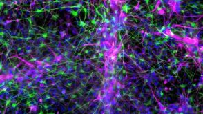 实验方案How to Co-Culture Astrocytes and NGN2 mRNA-Driven Induced Forebrain Neurons Derived from Human Pluripotent Stem Cells

 沪公网安备31010102008431号