Zeng J and Wang S (JAN 2014)
Stem cells translational medicine 3 1 69--80
Human dendritic cells derived from embryonic stem cells stably modified with CD1d efficiently stimulate antitumor invariant natural killer T cell response.
Invariant natural killer T (iNKT) cells are a unique lymphocyte subpopulation that mediates antitumor activities upon activation. A current strategy to harness iNKT cells for cancer treatment is endogenous iNKT cell activation using patient-derived dendritic cells (DCs). However,the limited number and functional defects of patient DCs are still the major challenges for this therapeutic approach. In this study,we investigated whether human embryonic stem cells (hESCs) with an ectopically expressed CD1d gene could be exploited to address this issue. Using a lentivector carrying an optimized expression cassette,we generated stably modified hESC lines that consistently overexpressed CD1d. These modified hESC lines were able to differentiate into DCs as efficiently as the parental line. Most importantly,more than 50% of such derived DCs were CD1d+. These CD1d-overexpressing DCs were more efficient in inducing iNKT cell response than those without modification,and their ability was comparable to that of DCs generated from monocytes of healthy donors. The iNKT cells expanded by the CD1d-overexpressing DCs were functional,as demonstrated by their ability to lyse iNKT cell-sensitive glioma cells. Therefore,hESCs stably modified with the CD1d gene may serve as a convenient,unlimited,and competent DC source for iNKT cell-based cancer immunotherapy.
View Publication
产品号#:
05850
05857
05870
05875
09600
09650
70024
70024.1
85850
85857
85870
85875
产品名:
StemSpan™ SFEM
StemSpan™ SFEM
冻存的人外周血Pan T细胞
冻存的人外周血Pan T细胞
mTeSR™1
mTeSR™1
Yang W-T and Zheng P-S (FEB 2014)
PloS one 9 2 e88827
Promoter hypermethylation of KLF4 inactivates its tumor suppressor function in cervical carcinogenesis.
OBJECTIVE The KLF4 gene has been shown to be inactivated in cervical carcinogenesis as a tumor suppressor. However,the mechanism of KLF4 silencing in cervical carcinomas has not yet been identified. DNA methylation plays a key role in stable suppression of gene expression. METHODS The methylation status of the KLF4 promoter CpG islands was analyzed by bisulfite sequencing (BSQ) in tissues of normal cervix and cervical cancer. KLF4 gene expression was detected by RT-PCR,immunohistochemistry and western blot. KLF4 promoter methylation in cervical cancer cell line was determined by BSQ and methylation-specific polymerase chain reaction (MS-PCR). Cell proliferation ability was detected by cell growth curve and MTT assay. RESULTS The methylated allele was found in 41.90% of 24 cervical cancer tissues but only in 11.11% of 11 normal cervix tissues (Ptextless0.005). KLF4 mRNA levels were significantly reduced in cervical cancer tissues compared with normal cervix tissues (Ptextless0.01) and KLF4 mRNA expression showed a significant negative correlation with the promoter hypermethylation (r = -0.486,P = 0.003). Cervical cancer cell lines also showed a significant negative correlation between KLF4 expression and hypermethylation. After treatment with the demethylating agent 5-Azacytidine (5-Aza),the expression of KLF4 in the cervical cancer cell lines at both mRNA and protein levels was drastically increased,the cell proliferation ability was inhibited and the chemosensitivity for cisplatin was significantly increased. CONCLUSION KLF4 gene is inactivated by methylation-induced silencing mechanisms in a large subset of cervical carcinomas and KLF4 promoter hypermethylation inactivates the gene's function as a tumor suppressor in cervical carcinogenesis.
View Publication
D. Duluc et al. ( 2014)
The Journal of Immunology 192 5776-88
Induction and activation of human Th17 by targeting antigens to dendritic cells via dectin-1
Recent compelling evidence indicates that Th17 confer host immunity against a variety of microbes,including extracellular and intracellular pathogens. Therefore,understanding mechanisms for the induction and activation of Ag-specific Th17 is important for the rational design of vaccines against pathogens. To study this,we employed an in vitro system in which influenza hemagglutinin (HA) 1 was delivered to dendritic cells (DCs) via Dectin-1 using anti-human Dectin-1 (hDectin-1)-HA1 recombinant fusion proteins. We found that healthy individuals maintained broad ranges of HA1-specific memory Th17 that were efficiently activated by DCs targeted with anti-hDectin-1-HA1. Nonetheless,these DCs were not able to induce a significant level of HA1-specific Th17 responses even in the presence of the Th17-promoting cytokines IL-1? and IL-6. We further found that the induction of surface IL-1R1 expression by signals via TCRs and common ?-chain receptors was essential for naive CD4(+) T cell differentiation into HA1-specific Th17. This process was dependent on MyD88,but not IL-1R-associated kinase 1/4. Thus,interruptions in STAT3 or MyD88 signaling led to substantially diminished HA1-specific Th17 induction. Taken together,the de novo generation of pathogen-specific human Th17 requires complex,but complementary,actions of multiple signals. Data from this study will help us design a new and effective vaccine strategy that can promote Th17-mediated immunity against microbial pathogens.
View Publication
产品号#:
19052
19052RF
产品名:
EasySep™人CD4+ T细胞富集试剂盒
RoboSep™ 人CD4+ T细胞富集试剂盒含滤芯吸头
S. Gupta et al. ( 2018)
Immunity & ageing : I & A 15 2
Molecular changes associated with increased TNF-?-induced apoptotis in naive (TN) and central memory (TCM) CD8+ T cells in aged humans.
Background Progressive T cell decline in aged humans is associated with a deficiency of naive (TN) and central memory (TCM) T cells. We have previously reported increased tumor necrosis factor-? (TNF-?)-induced apoptosis in TN and TCM T cells in aged humans; however,the molecular basis of increased apoptosis remains to be defined. Since expression of TNF receptors (TNFRs) was reported to be comparable in young and aged,we investigated signaling events downstream of TNFRs to understand the molecular basis of increased TNF-?-induced apoptosis in aged TN and TCM CD8+ cells. Results The expression of TRAF-2 and RIP,phosphorylation of JNK,IKK?/?,and I?B?,and activation of NF-?B activation were significantly decreased in TN and TCM CD8+ cells from aged subjects as compared to young controls. Furthermore,expression of A20,Bcl-xL,cIAP1,and FLIP-L and FLIP-S was significantly decreased in TN and TCM CD8+ cells from aged subjects. Conclusions These data demonstrate that an impaired expression/function of molecules downstream TNFR signaling pathway that confer survival signals contribute to increased apoptosis of TN and TCM CD8+ cells in aged humans.
View Publication

 EasySep™小鼠TIL(CD45)正选试剂盒


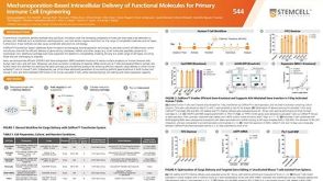 科学海报Mechanoporation-Based Intracellular Delivery of Functional Molecules for Primary Immune Cell Engineering


 沪公网安备31010102008431号