Ohoka Y et al. (JAN 2011)
Journal of immunology (Baltimore,Md. : 1950) 186 2 733--44
Retinoic acid-induced CCR9 expression requires transient TCR stimulation and cooperativity between NFATc2 and the retinoic acid receptor/retinoid X receptor complex.
Retinoic acid (RA) imprints gut-homing specificity on T cells upon activation by inducing the expression of chemokine receptor CCR9 and integrin α4β7. CCR9 expression seemed to be more highly dependent on RA than was the α4β7 expression,but its molecular mechanism remained unclear. In this article,we show that NFAT isoforms NFATc1 and NFATc2 directly interact with RA receptor (RAR) and retinoid X receptor (RXR) but play differential roles in RA-induced CCR9 expression on murine naive CD4(+) T cells. TCR stimulation for 6-24 h was required for the acquisition of responsiveness to RA and induced activation of NFATc1 and NFATc2. However,RA failed to induce CCR9 expression as long as TCR stimulation continued. After terminating TCR stimulation or adding cyclosporin A to the culture,Ccr9 gene transcription was induced,accompanied by inactivation of NFATc1 and sustained activation of NFATc2. Reporter and DNA-affinity precipitation assays demonstrated that the binding of NFATc2 to two NFAT-binding sites and that of the RAR/RXR complex to an RA response element half-site in the 5'-flanking region of the mouse Ccr9 gene were critical for RA-induced promoter activity. NFATc2 directly bound to RARα and RXRα,and it enhanced the binding of RARα to the RA response element half-site. NFATc1 also bound to the NFAT-binding sites and directly to RARα and RXRα,but it inhibited the NFATc2-dependent promoter activity. These results suggest that the cooperativity between NFATc2 and the RAR/RXR complex is essential for CCR9 expression on T cells and that NFATc1 interferes with the action of NFATc2.
View Publication
产品号#:
19752
19752RF
产品名:
Mkhikian H et al. (JAN 2011)
Nature communications 2 334
Genetics and the environment converge to dysregulate N-glycosylation in multiple sclerosis.
How environmental factors combine with genetic risk at the molecular level to promote complex trait diseases such as multiple sclerosis (MS) is largely unknown. In mice,N-glycan branching by the Golgi enzymes Mgat1 and/or Mgat5 prevents T cell hyperactivity,cytotoxic T-lymphocyte antigen 4 (CTLA-4) endocytosis,spontaneous inflammatory demyelination and neurodegeneration,the latter pathologies characteristic of MS. Here we show that MS risk modulators converge to alter N-glycosylation and/or CTLA-4 surface retention conditional on metabolism and vitamin D(3),including genetic variants in interleukin-7 receptor-α (IL7RA*C),interleukin-2 receptor-α (IL2RA*T),MGAT1 (IV(A)V(T-T)) and CTLA-4 (Thr17Ala). Downregulation of Mgat1 by IL7RA*C and IL2RA*T is opposed by MGAT1 (IV(A)V(T-T)) and vitamin D(3),optimizing branching and mitigating MS risk when combined with enhanced CTLA-4 N-glycosylation by CTLA-4 Thr17. Our data suggest a molecular mechanism in MS whereby multiple environmental and genetic inputs lead to dysregulation of a final common pathway,namely N-glycosylation.
View Publication
产品号#:
15021
15061
产品名:
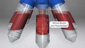
RosetteSep™人T细胞富集抗体混合物
RosetteSep™人T细胞富集抗体混合物
Decot V et al. (JAN 2008)
Bio-medical materials and engineering 18 1 Suppl S19--26
Chimerism analysis following nonmyeloablative stem cell transplantation using a new cell subset separation method: Robosep.
Chimerism analysis has become an important tool to manage patients in the peri-transplant period of allogenic stem cell transplantation. During this period,cells of donor and host origin can coexist and increasing proportion of cells of host origin is considered as a recurrence of the underlying disease. We currently performed chimerism analysis on separate peripheral blood cell subsets,lymphocytes and granulocytes. To improve our isolation method,a new automated device from Stem Cell Technology Roboseptrade mark was tested and compared to our manual separation technique. The results obtained on T cell purification showed an improvement of the purity (98.42% with Robosep vs. 92.42% with the manual technique Rosettesep) and of the recovery (63.43% with Robosep and 38% with Rosettesep). The results were significantly improved on patient samples with less than 10% CD3 positive cells (purity: 90% vs. 44.44%; recovery: 73.79% vs. 43.98%). Granulocytes separation was based on CD15 expression. The results showed an improvement of the purity with Robosep (96.90% vs. 86.20% with the manual technique Polymorphprep) but the recovery was impaired (35.2% vs. 52.30%). Using a myeloid (CD66/CD33) cocktail,recovery was improved with the Robosep device (64.04% with the myeloid cocktail vs. 22.4% with the CD15 cocktail). Our data demonstrated that Robosep allowed a performant cell purification in the early period post-transplantation even for populations representing less than 10% of the peripheral blood cells.
View Publication
产品号#:
19051
19051RF
21000
20119
20155
18681
18681RF
产品名:
EasySep™人T细胞富集试剂盒
RoboSep™ 人T细胞富集试剂盒含滤芯吸头
RoboSep™- S
RoboSep™ 吸头组件抛光剂
RoboSep™分选管套装(9个塑料管)
Strainic MG et al. (MAR 2008)
Immunity 28 3 425--35
Locally produced complement fragments C5a and C3a provide both costimulatory and survival signals to naive CD4+ T cells.
Costimulatory signals are critical to T cell activation,but how their effects are mediated remains incompletely characterized. Here,we demonstrate that locally produced C5a and C3a anaphylatoxins interacting with their G protein-coupled receptors (GPCRs),C5aR and C3aR,on APCs and T cells both upstream and downstream of CD28 and CD40L signaling are integrally involved in T cell proliferation and differentiation. Disabling these interactions reduced MHC class II and costimulatory-molecule expression and dramatically diminished T cell responses. Importantly,impaired T cell activation by Cd80-/-Cd86-/- and Cd40-/- APCs was reconstituted by added C5a or C3a. C5aR and C3aR mediated their effects via PI-3 kinase-gamma-dependent AKT phosphorylation,providing a link between GPCR signaling,CD28 costimulation,and T cell survival. These local paracrine and autocrine interactions thus operate constitutively in naive T cells to maintain viability,and their amplification by cognate APC partners thus is critical to T cell costimulation.
View Publication
Ichikawa S et al. (MAY 2011)
Journal of immunology (Baltimore,Md. : 1950) 186 10 5549--55
Hepatic stellate cells function as regulatory bystanders.
Regulatory T cells (Tregs) contribute significantly to the tolerogenic nature of the liver. The mechanisms,however,underlying liver-associated Treg induction are still elusive. We recently identified the vitamin A metabolite,retinoic acid (RA),as a key controller that promotes TGF-β-dependent Foxp3(+) Treg induction but inhibits TGF-β-driven Th17 differentiation. To investigate whether the RA producing hepatic stellate cells (HSC) are part of the liver tolerance mechanism,we investigated the ability of HSC to function as regulatory APC. Different from previous reports,we found that highly purified HSC did not express costimulatory molecules and only upregulated MHC class II after in vitro culture in the presence of exogenous IFN-γ. Consistent with an insufficient APC function,HSC failed to stimulate naive OT-II TCR transgenic CD4(+) T cells and only moderately stimulated α-galactosylceramide-primed invariant NKT cells. In contrast,HSC functioned as regulatory bystanders and promoted enhanced Foxp3 induction by OT-II TCR transgenic T cells primed by spleen dendritic cells,whereas they greatly inhibited the Th17 differentiation. Furthermore,the regulatory bystander capacity of the HSC was completely dependent on their ability to produce RA. Our data thus suggest that HSC can function as regulatory bystanders,and therefore,by promoting Tregs and suppressing Th17 differentiation,they might represent key players in the mechanism that drives liver-induced tolerance.
View Publication
产品号#:
01700
01705
01701
01702
19755
产品名:
ALDEFLUOR™ 试剂盒
ALDEFLUOR™ DEAB试剂, 1.5 mM, 1 mL
ALDEFLUOR™检测缓冲液
Li H et al. (AUG 2010)
Blood 116 7 1060--9
Repression of Id2 expression by Gfi-1 is required for B-cell and myeloid development.
The development of mature blood cells from hematopoietic stem cells requires coordinated activities of transcriptional networks. Transcriptional repressor growth factor independence 1 (Gfi-1) is required for the development of B cells,T cells,neutrophils,and for the maintenance of hematopoietic stem cell function. However,the mechanisms by which Gfi-1 regulates hematopoiesis and how Gfi-1 integrates into transcriptional networks remain unclear. Here,we provide evidence that Id2 is a transcriptional target of Gfi-1,and repression of Id2 by Gfi-1 is required for B-cell and myeloid development. Gfi-1 binds to 3 conserved regions in the Id2 promoter and represses Id2 promoter activity in transient reporter assays. Increased Id2 expression was observed in multipotent progenitors,myeloid progenitors,T-cell progenitors,and B-cell progenitors in Gfi-1(-/-) mice. Knockdown of Id2 expression or heterozygosity at the Id2 locus partially rescues the B-cell and myeloid development but not the T-cell development in Gfi-1(-/-) mice. These studies demonstrate a role of Id2 in mediating Gfi-1 functions in B-cell and myeloid development and provide a direct link between Gfi-1 and the B-cell transcriptional network by its ability to repress Id2 expression.
View Publication

 EasySep™小鼠TIL(CD45)正选试剂盒


 沪公网安备31010102008431号