S. Gupta et al. ( 2018)
Immunity & ageing : I & A 15 2
Molecular changes associated with increased TNF-?-induced apoptotis in naive (TN) and central memory (TCM) CD8+ T cells in aged humans.
Background Progressive T cell decline in aged humans is associated with a deficiency of naive (TN) and central memory (TCM) T cells. We have previously reported increased tumor necrosis factor-? (TNF-?)-induced apoptosis in TN and TCM T cells in aged humans; however,the molecular basis of increased apoptosis remains to be defined. Since expression of TNF receptors (TNFRs) was reported to be comparable in young and aged,we investigated signaling events downstream of TNFRs to understand the molecular basis of increased TNF-?-induced apoptosis in aged TN and TCM CD8+ cells. Results The expression of TRAF-2 and RIP,phosphorylation of JNK,IKK?/?,and I?B?,and activation of NF-?B activation were significantly decreased in TN and TCM CD8+ cells from aged subjects as compared to young controls. Furthermore,expression of A20,Bcl-xL,cIAP1,and FLIP-L and FLIP-S was significantly decreased in TN and TCM CD8+ cells from aged subjects. Conclusions These data demonstrate that an impaired expression/function of molecules downstream TNFR signaling pathway that confer survival signals contribute to increased apoptosis of TN and TCM CD8+ cells in aged humans.
View Publication
Interleukins 7 and 15 Maintain Human T Cell Proliferative Capacity through STAT5 Signaling.
T lymphocytes require signals from self-peptides and cytokines,most notably interleukins 7 and 15 (IL-7,IL-15),for survival. While mouse T cells die rapidly if IL-7 or IL-15 is withdrawn,human T cells can survive prolonged withdrawal of IL-7 and IL-15. Here we show that IL-7 and IL-15 are required to maintain human T cell proliferative capacity through the STAT5 signaling pathway. T cells from humanized mice proliferate better if stimulated in the presence of human IL-7 or IL-15 or if T cells are exposed to human IL-7 or IL-15 in mice. Freshly isolated T cells from human peripheral blood lose proliferative capacity if cultured for 24 hours in the absence of IL-7 or IL-15. We further show that phosphorylation of STAT5 correlates with proliferation and inhibition of STAT5 reduces proliferation. These results reveal a novel role of IL-7 and IL-15 in maintaining human T cell function,provide an explanation for T cell dysfunction in humanized mice,and have significant implications for in vitro studies with human T cells.
View Publication
产品号#:
17951
17951RF
19851
19851RF
15624
15664
100-0695
产品名:
EasySep™人T细胞分选试剂盒
RoboSep™ 人T细胞分选试剂盒
EasySep™小鼠T细胞分选试剂盒
RoboSep™ 小鼠T细胞分选试剂盒
RosetteSep™人粒细胞去除抗体混合物
RosetteSep™人粒细胞去除抗体混合物
EasySep™人T细胞分选试剂盒
von Bonin A et al. (JAN 2011)
Experimental dermatology 20 1 41--7
Inhibition of the IL-2-inducible tyrosine kinase (Itk) activity: a new concept for the therapy of inflammatory skin diseases.
T-cell-mediated processes play an essential role in the pathogenesis of several inflammatory skin diseases such as atopic dermatitis,allergic contact dermatitis and psoriasis. The aim of this study was to investigate the role of the IL-2-inducible tyrosine kinase (Itk),an enzyme acting downstream of the T-cell receptor (TCR),in T-cell-dependent skin inflammation using three approaches. Itk knockout mice display significantly reduced inflammatory symptoms in mouse models of acute and subacute contact hypersensitivity (CHS) reactions. Systemic administration of a novel small molecule Itk inhibitor,Compound 44,created by chemical optimization of an initial high-throughput screening hit,inhibited Itk's activity with an IC50 in the nanomolar range. Compound 44 substantially reduced proinflammatory immune responses in vitro and in vivo after systemic administration in two acute CHS models. In addition,our data reveal that human Itk,comparable to its murine homologue,is expressed mainly in T cells and is increased in lesional skin from patients with atopic dermatitis and allergic contact dermatitis. Finally,silencing of Itk by RNA interference in primary human T cells efficiently blocks TCR-induced lymphokine secretion. In conclusion,Itk represents an interesting new target for the therapy of T-cell-mediated inflammatory skin diseases.
View Publication
产品号#:
15021
15061
产品名:
RosetteSep™人T细胞富集抗体混合物
RosetteSep™人T细胞富集抗体混合物
L. L. Lu et al. ( 2019)
Nature medicine 25 6 977--987
IFN-gamma-independent immune markers of Mycobacterium tuberculosis exposure.
Exposure to Mycobacterium tuberculosis (Mtb) results in heterogeneous clinical outcomes including primary progressive tuberculosis and latent Mtb infection (LTBI). Mtb infection is identified using the tuberculin skin test and interferon-gamma (IFN-gamma) release assay IGRA,and a positive result may prompt chemoprophylaxis to prevent progression to tuberculosis. In the present study,we report on a cohort of Ugandan individuals who were household contacts of patients with TB. These individuals were highly exposed to Mtb but tested negative disease by IFN-gamma release assay and tuberculin skin test,'resisting' development of classic LTBI. We show that 'resisters' possess IgM,class-switched IgG antibody responses and non-IFN-gamma T cell responses to the Mtb-specific proteins ESAT6 and CFP10,immunologic evidence of exposure to Mtb. Compared to subjects with classic LTBI,'resisters' display enhanced antibody avidity and distinct Mtb-specific IgG Fc profiles. These data reveal a distinctive adaptive immune profile among Mtb-exposed subjects,supporting an expanded definition of the host response to Mtb exposure,with implications for public health and the design of clinical trials.
View Publication
A. A. Titov et al. (jul 2019)
Journal of immunology (Baltimore,Md. : 1950) 203 2 338--348
Metformin Inhibits the Type 1 IFN Response in Human CD4+ T Cells.
In systemic lupus erythematosus,defective clearance of apoptotic debris and activation of innate cells result in a chronically activated type 1 IFN response,which can be measured in PBMCs of most patients. Metformin,a widely used prescription drug for Type 2 diabetes,has a therapeutic effect in several mouse models of lupus through mechanisms involving inhibition of oxidative phosphorylation and a decrease in CD4+ T cell activation. In this study,we report that in CD4+ T cells from human healthy controls and human systemic lupus erythematosus patients,metformin inhibits the transcription of IFN-stimulated genes (ISGs) after IFN-alpha treatment. Accordingly,metformin inhibited the phosphorylation of pSTAT1 (Y701) and its binding to IFN-stimulated response elements that control ISG expression. These effects were independent of AMPK activation or mTORC1 inhibition but were replicated using inhibitors of the electron transport chain respiratory complexes I,III,and IV. This indicates that mitochondrial respiration is required for ISG expression in CD4+ T cells and provides a novel mechanism by which metformin may exert a therapeutic effect in autoimmune diseases.
View Publication
Ohoka Y et al. (JAN 2011)
Journal of immunology (Baltimore,Md. : 1950) 186 2 733--44
Retinoic acid-induced CCR9 expression requires transient TCR stimulation and cooperativity between NFATc2 and the retinoic acid receptor/retinoid X receptor complex.
Retinoic acid (RA) imprints gut-homing specificity on T cells upon activation by inducing the expression of chemokine receptor CCR9 and integrin α4β7. CCR9 expression seemed to be more highly dependent on RA than was the α4β7 expression,but its molecular mechanism remained unclear. In this article,we show that NFAT isoforms NFATc1 and NFATc2 directly interact with RA receptor (RAR) and retinoid X receptor (RXR) but play differential roles in RA-induced CCR9 expression on murine naive CD4(+) T cells. TCR stimulation for 6-24 h was required for the acquisition of responsiveness to RA and induced activation of NFATc1 and NFATc2. However,RA failed to induce CCR9 expression as long as TCR stimulation continued. After terminating TCR stimulation or adding cyclosporin A to the culture,Ccr9 gene transcription was induced,accompanied by inactivation of NFATc1 and sustained activation of NFATc2. Reporter and DNA-affinity precipitation assays demonstrated that the binding of NFATc2 to two NFAT-binding sites and that of the RAR/RXR complex to an RA response element half-site in the 5'-flanking region of the mouse Ccr9 gene were critical for RA-induced promoter activity. NFATc2 directly bound to RARα and RXRα,and it enhanced the binding of RARα to the RA response element half-site. NFATc1 also bound to the NFAT-binding sites and directly to RARα and RXRα,but it inhibited the NFATc2-dependent promoter activity. These results suggest that the cooperativity between NFATc2 and the RAR/RXR complex is essential for CCR9 expression on T cells and that NFATc1 interferes with the action of NFATc2.
View Publication
产品号#:
19752
19752RF
产品名:
Mkhikian H et al. (JAN 2011)
Nature communications 2 334
Genetics and the environment converge to dysregulate N-glycosylation in multiple sclerosis.
How environmental factors combine with genetic risk at the molecular level to promote complex trait diseases such as multiple sclerosis (MS) is largely unknown. In mice,N-glycan branching by the Golgi enzymes Mgat1 and/or Mgat5 prevents T cell hyperactivity,cytotoxic T-lymphocyte antigen 4 (CTLA-4) endocytosis,spontaneous inflammatory demyelination and neurodegeneration,the latter pathologies characteristic of MS. Here we show that MS risk modulators converge to alter N-glycosylation and/or CTLA-4 surface retention conditional on metabolism and vitamin D(3),including genetic variants in interleukin-7 receptor-α (IL7RA*C),interleukin-2 receptor-α (IL2RA*T),MGAT1 (IV(A)V(T-T)) and CTLA-4 (Thr17Ala). Downregulation of Mgat1 by IL7RA*C and IL2RA*T is opposed by MGAT1 (IV(A)V(T-T)) and vitamin D(3),optimizing branching and mitigating MS risk when combined with enhanced CTLA-4 N-glycosylation by CTLA-4 Thr17. Our data suggest a molecular mechanism in MS whereby multiple environmental and genetic inputs lead to dysregulation of a final common pathway,namely N-glycosylation.
View Publication

 EasySep™小鼠TIL(CD45)正选试剂盒


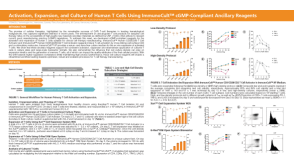 科学海报Activation, Expansion, and Culture of Human T Cells Using ImmunoCult™ cGMP-Compliant Ancillary Reagents


 沪公网安备31010102008431号