Zhao L et al. (SEP 2014)
Stem Cell Research 13 2 342--354
Heterelogous expression of mutated HLA-G decreases immunogenicity of human embryonic stem cells and their epidermal derivatives.
Human embryonic stem cells (hESCs) are capable of extensive self-renewal and expansion and can differentiate into any somatic tissue,making them useful for regenerative medicine applications. Allogeneic transplantation of hESC-derived tissues from results in immunological rejection absent adjunctive immunosuppression. The goal of our study was to generate a universal pluripotent stem cell source by nucleofecting a mutated human leukocyte antigen G (mHLA-G) gene into hESCs using the PiggyBac transposon. We successfully generated stable mHLA-G(EF1$\$)-hESC lines using chEF1$\$ system that stably expressed mHLA-G protein during prolonged undifferentiated proliferation andin differentiated embryoid bodies as well as teratomas. Morphology,karyotype,and telomerase activity of mHLA-G expressing hESC were normal. Immunofluorescence staining and flow cytometry analysis revealed persistent expression of pluripotent markers,OCT-3/4 and SSEA-4,in undifferentiated mHLA-G(EF1$\$)-hESC. Nucleofected hESC formed teratomas and when directed to differentiate into epidermal precursors,expressed high levels of mHLA-G and keratinocyte markers K14 and CD29. Natural killer cell cytotoxicity assays demonstrated a significant decrease in lysis of mHLA-G(EF1a)-hESC targets relative to control cells. Similar results were obtained with mHLA-G(EF1$\$)-hESC-derived epidermal progenitors (hEEP). One way mixed T lymphocyte reactions unveiled that mHLA-G(EF1a)-hESC and -hEEP restrained the proliferative activity of mixed T lymphocytes. We conclude that heterologous expression of mHLA-G decreases immunogenicity of hESCs and their epidermal differentiated derivatives.
View Publication
CD80 and PD-L2 define functionally distinct memory B cell subsets that are independent of antibody isotype
Memory B cells (MBCs) are long-lived sources of rapid,isotype-switched secondary antibody-forming cell (AFC) responses. Whether MBCs homogeneously retain the ability to self-renew and terminally differentiate or if these functions are compartmentalized into MBC subsets has remained unclear. It has been suggested that antibody isotype controls MBC differentiation upon restimulation. Here we demonstrate that subcategorizing MBCs on the basis of their expression of CD80 and PD-L2,independently of isotype,identified MBC subsets with distinct functions upon rechallenge. CD80(+)PD-L2(+) MBCs differentiated rapidly into AFCs but did not generate germinal centers (GCs); conversely,CD80(-)PD-L2(-) MBCs generated few early AFCs but robustly seeded GCs. The gene-expression patterns of the subsets supported both the identity and function of these distinct MBC types. Hence,the differentiation and regeneration of MBCs are compartmentalized.
View Publication
产品号#:
19752
19752RF
19754
19754RF
产品名:
Yang W-T and Zheng P-S (FEB 2014)
PloS one 9 2 e88827
Promoter hypermethylation of KLF4 inactivates its tumor suppressor function in cervical carcinogenesis.
OBJECTIVE The KLF4 gene has been shown to be inactivated in cervical carcinogenesis as a tumor suppressor. However,the mechanism of KLF4 silencing in cervical carcinomas has not yet been identified. DNA methylation plays a key role in stable suppression of gene expression. METHODS The methylation status of the KLF4 promoter CpG islands was analyzed by bisulfite sequencing (BSQ) in tissues of normal cervix and cervical cancer. KLF4 gene expression was detected by RT-PCR,immunohistochemistry and western blot. KLF4 promoter methylation in cervical cancer cell line was determined by BSQ and methylation-specific polymerase chain reaction (MS-PCR). Cell proliferation ability was detected by cell growth curve and MTT assay. RESULTS The methylated allele was found in 41.90% of 24 cervical cancer tissues but only in 11.11% of 11 normal cervix tissues (Ptextless0.005). KLF4 mRNA levels were significantly reduced in cervical cancer tissues compared with normal cervix tissues (Ptextless0.01) and KLF4 mRNA expression showed a significant negative correlation with the promoter hypermethylation (r = -0.486,P = 0.003). Cervical cancer cell lines also showed a significant negative correlation between KLF4 expression and hypermethylation. After treatment with the demethylating agent 5-Azacytidine (5-Aza),the expression of KLF4 in the cervical cancer cell lines at both mRNA and protein levels was drastically increased,the cell proliferation ability was inhibited and the chemosensitivity for cisplatin was significantly increased. CONCLUSION KLF4 gene is inactivated by methylation-induced silencing mechanisms in a large subset of cervical carcinomas and KLF4 promoter hypermethylation inactivates the gene's function as a tumor suppressor in cervical carcinogenesis.
View Publication
产品号#:
05850
05857
05870
05875
85850
85857
85870
85875
产品名:
mTeSR™1
mTeSR™1
Zeng J and Wang S (JAN 2014)
Stem cells translational medicine 3 1 69--80
Human dendritic cells derived from embryonic stem cells stably modified with CD1d efficiently stimulate antitumor invariant natural killer T cell response.
Invariant natural killer T (iNKT) cells are a unique lymphocyte subpopulation that mediates antitumor activities upon activation. A current strategy to harness iNKT cells for cancer treatment is endogenous iNKT cell activation using patient-derived dendritic cells (DCs). However,the limited number and functional defects of patient DCs are still the major challenges for this therapeutic approach. In this study,we investigated whether human embryonic stem cells (hESCs) with an ectopically expressed CD1d gene could be exploited to address this issue. Using a lentivector carrying an optimized expression cassette,we generated stably modified hESC lines that consistently overexpressed CD1d. These modified hESC lines were able to differentiate into DCs as efficiently as the parental line. Most importantly,more than 50% of such derived DCs were CD1d+. These CD1d-overexpressing DCs were more efficient in inducing iNKT cell response than those without modification,and their ability was comparable to that of DCs generated from monocytes of healthy donors. The iNKT cells expanded by the CD1d-overexpressing DCs were functional,as demonstrated by their ability to lyse iNKT cell-sensitive glioma cells. Therefore,hESCs stably modified with the CD1d gene may serve as a convenient,unlimited,and competent DC source for iNKT cell-based cancer immunotherapy.
View Publication
产品号#:
05850
05857
05870
05875
09600
09650
70024
70024.1
85850
85857
85870
85875
产品名:
StemSpan™ SFEM
StemSpan™ SFEM
冻存的人外周血Pan T细胞
冻存的人外周血Pan T细胞
mTeSR™1
mTeSR™1
Rosenzweig M et al. (APR 2001)
Blood 97 7 1951--9
Induction of cytotoxic T lymphocyte and antibody responses to enhanced green fluorescent protein following transplantation of transduced CD34(+) hematopoietic cells.
Genetic modification of hematopoietic stem cells often results in the expression of foreign proteins in pluripotent progenitor cells and their progeny. However,the potential for products of foreign genes introduced into hematopoietic stem cells to induce host immune responses is not well understood. Gene marking and induction of immune responses to enhanced green fluorescent protein (eGFP) were examined in rhesus macaques that underwent nonmyeloablative irradiation followed by infusions of CD34(+) bone marrow cells transduced with a retroviral vector expressing eGFP. CD34(+) cells were obtained from untreated animals or from animals treated with recombinant human granulocyte colony-stimulating factor (G-CSF) alone or G-CSF and recombinant human stem cell factor. Levels of eGFP-expressing cells detected by flow cytometry peaked at 0.1% to 0.5% of all leukocytes 1 to 4 weeks after transplantation. Proviral DNA was detected in 0% to 17% of bone marrow--derived colony-forming units at periods of 5 to 18 weeks after transplantation. However,5 of 6 animals studied demonstrated a vigorous eGFP-specific cytotoxic T lymphocyte (CTL) response that was associated with a loss of genetically modified cells in peripheral blood,as demonstrated by both flow cytometry and polymerase chain reaction. The eGFP-specific CTL responses were MHC-restricted,mediated by CD8(+) lymphocytes,and directed against multiple epitopes. eGFP-specific CTLs were able to efficiently lyse autologous CD34(+) cells expressing eGFP. Antibody responses to eGFP were detected in 3 of 6 animals. These data document the potential for foreign proteins expressed in CD34(+) hematopoietic cells and their progeny to induce antibody and CTL responses in the setting of a clinically applicable transplantation protocol. (Blood. 2001;97:1951-1959)
View Publication
产品号#:
09600
09650
产品名:
StemSpan™ SFEM
StemSpan™ SFEM
Cho SK et al. (AUG 1999)
Proceedings of the National Academy of Sciences of the United States of America 96 17 9797--802
Functional characterization of B lymphocytes generated in vitro from embryonic stem cells.
To study molecular events involved in B lymphocyte development and V(D)J rearrangement,we have established an efficient system for the differentiation of embryonic stem (ES) cells into mature Ig-secreting B lymphocytes. Here,we show that B lineage cells generated in vitro from ES cells are functionally analogous to normal fetal liver-derived or bone marrow-derived B lineage cells at three important developmental stages: first,they respond to Flt-3 ligand during an early lymphopoietic progenitor stage; second,they become targets for Abelson murine leukemia virus (A-MuLV) infection at a pre-B cell stage; third,they secrete Ig upon stimulation with lipopolysaccharide at a mature mitogen-responsive stage. Moreover,the ES cell-derived A-MuLV-transformed pre-B (EAB) cells are phenotypically and functionally indistinguishable from standard A-MuLV-transformed pre-B cells derived from infection of mouse fetal liver or bone marrow. Notably,EAB cells possess functional V(D)J recombinase activity. In particular,the generation of A-MuLV transformants from ES cells will provide an advantageous system to investigate genetic modifications that will help to elucidate molecular mechanisms in V(D)J recombination and in A-MuLV-mediated transformation.
View Publication
Human Immune Cytokines
Infographic of key cytokines for expansion, differentiation and characterization of major immune cell types
Keller G et al. (JAN 1993)
Molecular and cellular biology 13 1 473--86
Hematopoietic commitment during embryonic stem cell differentiation in culture.
We report that embryonic stem cells efficiently undergo differentiation in vitro to mesoderm and hematopoietic cells and that this in vitro system recapitulates days 6.5 to 7.5 of mouse hematopoietic development. Embryonic stem cells differentiated as embryoid bodies (EBs) develop erythroid precursors by day 4 of differentiation,and by day 6,more than 85% of EBs contain such cells. A comparative reverse transcriptase-mediated polymerase chain reaction profile of marker genes for primitive endoderm (collagen alpha IV) and mesoderm (Brachyury) indicates that both cell types are present in the developing EBs as well in normal embryos prior to the onset of hematopoiesis. GATA-1,GATA-3,and vav are expressed in both the EBs and embryos just prior to and/or during the early onset of hematopoiesis,indicating that they could play a role in the early stages of hematopoietic development both in vivo and in vitro. The initial stages of hematopoietic development within the EBs occur in the absence of added growth factors and are not significantly influenced by the addition of a broad spectrum of factors,including interleukin-3 (IL-3),IL-1,IL-6,IL-11,erythropoietin,and Kit ligand. At days 10 and 14 of differentiation,EB hematopoiesis is significantly enhanced by the addition of both Kit ligand and IL-11 to the cultures. Kinetic analysis indicates that hematopoietic precursors develop within the EBs in an ordered pattern. Precursors of the primitive erythroid lineage appear first,approximately 24 h before precursors of the macrophage and definitive erythroid lineages. Bipotential neutrophil/macrophage and multilineage precursors appear next,and precursors of the mast cell lineage develop last. The kinetics of precursor development,as well as the growth factor responsiveness of these early cells,is similar to that found in the yolk sac and early fetal liver,indicating that the onset of hematopoiesis within the EBs parallels that found in the embryo.
View Publication
产品号#:
06902
06952
00321
00322
00323
00324
00325
产品名:
Gilbert C et al. (JUL 2007)
Journal of virology 81 14 7672--82
Human immunodeficiency virus type 1 replication in dendritic cell-T-cell cocultures is increased upon incorporation of host LFA-1 due to higher levels of virus production in immature dendritic cells.
Dendritic cells (DCs) act as a portal for invasion by human immunodeficiency virus type-1 (HIV-1). Here,we investigated whether virion-incorporated host cell membrane proteins can affect virus replication in DC-T-cell cocultures. Using isogenic viruses either devoid of or bearing host-derived leukocyte function-associated antigen 1 (LFA-1),we showed that HIV-1 production is augmented when LFA-1-bearing virions are used compared to that for viral entities lacking this adhesion molecule. This phenomenon was observed in immature monocyte-derived DCs (IM-MDDCs) only and not in DCs displaying a mature phenotype. The increase is not due to higher virus production in responder CD4(+) T cells but rather is linked with a more important productive infection of IM-MDDCs. We provided evidence that virus-associated host LFA-1 molecules do not affect a late event in the HIV-1 life cycle but rather exert an effect on an early step in virus replication. We demonstrated that the enhancement of productive infection of IM-MDDCs that is conferred by virus-anchored host LFA-1 involves the protein kinase A (PKA) and PKC signal transduction pathways. The biological significance of this phenomenon was established by performing experiments with virus stocks produced in primary human cells and anti-LFA-1 antibodies. Together,our results indicate that the association between some virus-bound host proteins and their natural cognate ligands can modulate de novo HIV-1 production by IM-MDDCs. Therefore,the additional interactions between virus-bound host cell membrane constituents and counter receptors on the surfaces of DCs can influence HIV-1 replication in IM-MDDC-T-cell cocultures.
View Publication
产品号#:
18058
18058RF
19052
19052RF
产品名:
EasySep™人CD4+ T细胞富集试剂盒
RoboSep™ 人CD4+ T细胞富集试剂盒含滤芯吸头
Nair S et al. (JAN 2007)
Cancer research 67 1 371--80
Vaccination against the forkhead family transcription factor Foxp3 enhances tumor immunity.
Depletion of CD4+CD25+ regulatory T cells (Treg) by treatment with alphaCD25 antibody synergizes with vaccination protocols to engender protective immunity in mice. The effectiveness of targeting CD25 to eliminate Treg is limited by the fact that CD25,the low-affinity interleukin-2 receptor,is up-regulated on conventional T cells. At present,foxp3 is the only product known to be exclusively expressed in Treg of mice. However,foxp3 is not expressed on the cell surface and hence cannot be targeted with antibodies. In this study,we tested the hypothesis that vaccination of mice against foxp3,a self-antigen expressed also in the thymus,is capable of stimulating foxp3-specific CTL that will cause the depletion of Treg and enhanced antitumor immunity. Vaccination of mice with foxp3 mRNA-transfected dendritic cells elicited a robust foxp3-specific CTL response and potentiated vaccine-induced protective immunity comparably with that of alphaCD25 antibody administration. In contrast to alphaCD25 antibody treatment,repeated foxp3 vaccination did not interfere with vaccine-induced protective immunity. Importantly,foxp3 vaccination led to the preferential depletion of foxp3-expressing Treg in the tumor but not in the periphery,whereas alphaCD25 antibody treatment led to depletion of Treg in both the tumor and the periphery. Targeting foxp3 by vaccination offers a specific and simpler protocol for the prolonged control of Treg that may be associated with reduced risk of autoimmunity,introducing an approach whereby specific depletion of cells is not limited to targeting products expressed on the cell surface.
View Publication

 EasySep™小鼠TIL(CD45)正选试剂盒


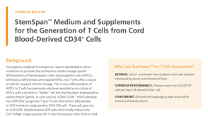 技术公告StemSpan™ Medium and Supplements for the Generation of T Cells from Cord Blood-Derived CD34+ Cells
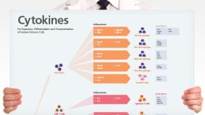 挂图Human Immune Cytokines Infographic of key cytokines for expansion, differentiation and characterization of major immune cell types

 沪公网安备31010102008431号