R. Lorenzetti et al. (jul 2019)
Journal of autoimmunity 101 145--152
Abatacept modulates CD80 and CD86 expression and memory formation in human B-cells.
BACKGROUND Cytotoxic T lymphocyte antigen-4 (CTLA-4) limits T-cell activation and is expressed on T-regulatory cells. Human CTLA-4 deficiency results in severe immune dysregulation. Abatacept (CTLA-4 Ig) is approved for the treatment of rheumatoid arthritis (RA) and its mechanism of action is attributed to effects on T-cells. It is known that CTLA-4 modulates the expression of its ligands CD80 and CD86 on antigen presenting cells (APC) by transendocytosis. As B-cells express CD80/CD86 and function as APC,we hypothesize that B-cells are a direct target of abatacept. OBJECTIVES To investigate direct effects of abatacept on human B-lymphocytes in vitro and in RA patients. METHODS The effect of abatacept on healthy donor B-cells' phenotype,activation and CD80/CD86 expression was studied in vitro. Nine abatacept-treated RA patients were studied. Seven of these were followed up to 24 months,and two up to 12 months only and treatment response,immunoglobulins,ACPA,RF concentrations,B-cell phenotype and ACPA-specific switched memory B-cell frequency were assessed. RESULTS B-cell development was unaffected by abatacept. Abatacept treatment resulted in a dose-dependent decrease of CD80/CD86 expression on B-cells in vitro,which was due to dynamin-dependent internalization. RA patients treated with abatacept showed a progressive decrease in plasmablasts and serum IgG. While ACPA-titers only moderately declined,the frequency of ACPA-specific switched memory B-cells significantly decreased. CONCLUSIONS Abatacept directly targets B-cells by reducing CD80/CD86 expression. Impairment of antigen presentation and T-cell activation may result in altered B-cell selection,providing a new therapeutic mechanism and a base for abatacept use in B-cell mediated autoimmunity.
View Publication
产品号#:
17954
17954RF
100-0971
产品名:
EasySep™人B细胞分选试剂盒
RoboSep™ 人B细胞分选试剂盒
EasySep™人B细胞分离试剂盒
P. Petrov et al. (mar 2019)
Scientific reports 9 1 4155
Computational analysis of the evolutionarily conserved Missing In Metastasis/Metastasis Suppressor 1 gene predicts novel interactions, regulatory regions and transcriptional control.
Missing in Metastasis (MIM),or Metastasis Suppressor 1 (MTSS1),is a highly conserved protein,which links the plasma membrane to the actin cytoskeleton. MIM has been implicated in various cancers,however,its modes of action remain largely enigmatic. Here,we performed an extensive in silico characterisation of MIM to gain better understanding of its function. We detected previously unappreciated functional motifs including adaptor protein (AP) complex interaction site and a C-helix,pointing to a role in endocytosis and regulation of actin dynamics,respectively. We also identified new functional regions,characterised with phosphorylation sites or distinct hydrophilic properties. Strong negative selection during evolution,yielding high conservation of MIM,has been combined with positive selection at key sites. Interestingly,our analysis of intra-molecular co-evolution revealed potential regulatory hotspots that coincided with reduced potentially pathogenic polymorphisms. We explored databases for the mutations and expression levels of MIM in cancer. Experimentally,we focused on chronic lymphocytic leukaemia (CLL),where MIM showed high overall expression,however,downregulation on poor prognosis samples. Finally,we propose strong conservation of MTSS1 also on the transcriptional level and predict novel transcriptional regulators. Our data highlight important targets for future studies on the role of MIM in different tissues and cancers.
View Publication
产品号#:
15024
15064
产品名:
RosetteSep™人B细胞富集抗体混合物
RosetteSep™人B细胞富集抗体混合物
Arendt BK et al. (SEP 2008)
Blood 112 5 1931--41
Biologic and genetic characterization of the novel amyloidogenic lambda light chain-secreting human cell lines, ALMC-1 and ALMC-2.
Primary systemic amyloidosis (AL) is a rare monoclonal plasma cell (PC) disorder characterized by the deposition of misfolded immunoglobulin (Ig) light chains (LC) in vital organs throughout the body. To our knowledge,no cell lines have ever been established from AL patients. Here we describe the establishment of the ALMC-1 and ALMC-2 cell lines from an AL patient. Both cell lines exhibit a PC phenotype and display cytokine-dependent growth. Using a comprehensive genetic approach,we established the genetic relationship between the cell lines and the primary patient cells,and we were also able to identify new genetic changes accompanying tumor progression that may explain the natural history of this patient's disease. Importantly,we demonstrate that free lambda LC secreted by both cell lines contained a beta structure and formed amyloid fibrils. Despite absolute Ig LC variable gene sequence identity,the proteins show differences in amyloid formation kinetics that are abolished by the presence of Na(2)SO(4). The formation of amyloid fibrils from these naturally secreting human LC cell lines is unprecedented. Moreover,these cell lines will provide an invaluable tool to better understand AL,from the combined perspectives of amyloidogenic protein structure and amyloid formation,genetics,and cell biology.
View Publication
产品号#:
18357
18357RF
21000
20119
20155
18387
18387RF
产品名:
RoboSep™- S
RoboSep™ 吸头组件抛光剂
RoboSep™分选管套装(9个塑料管)
Le Y et al. (MAR 2005)
Journal of immunology (Baltimore,Md. : 1950) 174 5 2582--90
CXC chemokine ligand 12-induced focal adhesion kinase activation and segregation into membrane domains is modulated by regulator of G protein signaling 1 in pro-B cells.
CXCL12-induced chemotaxis and adhesion to VCAM-1 decrease as B cells differentiate in the bone marrow. However,the mechanisms that regulate CXCL12/CXCR4-mediated signaling are poorly understood. We report that after CXCL12 stimulation of progenitor B cells,focal adhesion kinase (FAK) and PI3K are inducibly recruited to raft-associated membrane domains. After CXCL12 stimulation,phosphorylated FAK is also localized in membrane domains. The CXCL12/CXCR4-FAK pathway is membrane cholesterol dependent and impaired by metabolic inhibitors of G(i),Src family,and the GTPase-activating protein,regulator of G protein signaling 1 (RGS1). In the bone marrow,RGS1 mRNA expression is low in progenitor B cells and high in mature B cells,implying developmental regulation of CXCL12/CXCR4 signaling by RGS1. CXCL12-induced chemotaxis and adhesion are impaired when FAK recruitment and phosphorylation are inhibited by either membrane cholesterol depletion or overexpression of RGS1 in progenitor B cells. We conclude that the recruitment of signaling molecules to specific membrane domains plays an important role in CXCL12/CXCR4-induced cellular responses.
View Publication
产品号#:
产品名:
Balakrishnan K et al. (OCT 2006)
Blood 108 7 2392--8
Forodesine, an inhibitor of purine nucleoside phosphorylase, induces apoptosis in chronic lymphocytic leukemia cells.
Purine nucleoside phosphorylase (PNP) deficiency in humans results in T lymphocytopenia. Forodesine,a potent inhibitor of PNP,was designed based on the transition-state structure stabilized by the enzyme. Previous studies established that forodesine in the presence of deoxyguanosine (dGuo) inhibits the proliferation of T lymphocytes. A phase 1 clinical trial of forodesine in T-cell malignancies demonstrated significant antileukemic activity with an increase in intracellular dGuo triphosphate (dGTP). High accumulation of dGTP in T cells may be dependent on the levels of deoxynucleoside kinases. Because B-cell chronic lymphocytic leukemia (B-CLL) cells have high activity of deoxycytidine kinase (dCK),we hypothesized that these lymphocytes would respond to forodesine. This postulate was tested in primary lymphocytes during in vitro investigations. Lymphocytes from 12 patients with CLL were incubated with forodesine and dGuo. These CLL cells showed a wide variation in the accumulation of intracellular dGTP without any effect on other deoxynucleotides. This was associated with DNA damage-induced p53 stabilization,phosphorylation of p53 at Ser15,and activation of p21. The dGTP accumulation was related to induction of apoptosis measured by caspase activation,changes in mitochondrial membrane potential,and PARP cleavage. Based on these data,a phase 2 clinical trial of forodesine has been initiated for CLL patients.
View Publication
产品号#:
19051
19051RF
19054
19054RF
产品名:
EasySep™人T细胞富集试剂盒
RoboSep™ 人T细胞富集试剂盒含滤芯吸头
EasySep™人B细胞富集试剂盒
RoboSep™ 人B细胞富集试剂盒含滤芯吸头
Kharas MG et al. (JAN 2007)
Blood 109 2 747--55
KLF4 suppresses transformation of pre-B cells by ABL oncogenes.
Genes that are strongly repressed after B-cell activation are candidates for being inactivated,mutated,or repressed in B-cell malignancies. Krüppel-like factor 4 (Klf4),a gene down-regulated in activated murine B cells,is expressed at low levels in several types of human B-cell lineage lymphomas and leukemias. The human KLF4 gene has been identified as a tumor suppressor gene in colon and gastric cancer; in concordance with this,overexpression of KLF4 can suppress proliferation in several epithelial cell types. Here we investigate the effects of KLF4 on pro/pre-B-cell transformation by v-Abl and BCR-ABL,oncogenes that cause leukemia in mice and humans. We show that overexpression of KLF4 induces arrest and apoptosis in the G1 phase of the cell cycle. KLF4-mediated death,but not cell-cycle arrest,can be rescued by Bcl-XL overexpression. Transformed pro/pre-B cells expressing KLF4 display increased expression of p21CIP and decreased expression of c-Myc and cyclin D2. Tetracycline-inducible expression of KLF4 in B-cell progenitors of transgenic mice blocks transformation by BCR-ABL and depletes leukemic pre-B cells in vivo. Collectively,our work identifies KLF4 as a putative tumor suppressor in B-cell malignancies.
View Publication
产品号#:
03630
产品名:
MethoCult™ M3630
Bourdeau A et al. (MAY 2007)
Blood 109 10 4220--8
TC-PTP-deficient bone marrow stromal cells fail to support normal B lymphopoiesis due to abnormal secretion of interferon-gamma.
The T-cell protein tyrosine phosphatase (TC-PTP) is a negative regulator of the Jak/Stat cytokine signaling pathway. Our study shows that the absence of TC-PTP leads to an early bone marrow B-cell deficiency characterized by hindered transition from the pre-B cell to immature B-cell stage. This phenotype is intrinsic to the B cells but most importantly due to bone marrow stroma abnormalities. We found that bone marrow stromal cells from TC-PTP(-/-) mice have the unique property of secreting 232-890 pg/mL IFN-gamma. These high levels of IFN-gamma result in 2-fold reduction in mitotic index on IL-7 stimulation of TC-PTP(-/-) pre-B cells and lower responsiveness of IL-7 receptor downstream Jak/Stat signaling molecules. Moreover,we noted constitutive phosphorylation of Stat1 in those pre-B cells and demonstrated that this was due to soluble IFN-gamma secreted by TC-PTP(-/-) bone marrow stromal cells. Interestingly,culturing murine early pre-B leukemic cells within a TC-PTP-deficient bone marrow stroma environment leads to a 40% increase in apoptosis in these malignant cells. Our results unraveled a new role for TC-PTP in normal B lymphopoiesis and suggest that modulation of bone marrow microenvironment is a potential therapeutic approach for selected B-cell leukemia.
View Publication
产品号#:
03630
03434
03444
产品名:
MethoCult™ M3630
MethoCult™ GF M3434
MethoCult™ GF M3434
Douglas KB et al. (JUL 2009)
Genes and immunity 10 5 457--69
Complement receptor 2 polymorphisms associated with systemic lupus erythematosus modulate alternative splicing.
Genetic factors influence susceptibility to systemic lupus erythematosus (SLE). A recent family-based analysis in Caucasian and Chinese populations provided evidence for association of single-nucleotide polymorphisms (SNPs) in the complement receptor 2 (CR2/CD21) gene with SLE. Here we confirmed this result in a case-control analysis of an independent European-derived population including 2084 patients with SLE and 2853 healthy controls. A haplotype formed by the minor alleles of three CR2 SNPs (rs1048971,rs17615,rs4308977) showed significant association with decreased risk of SLE (30.4% in cases vs 32.6% in controls,P=0.016,OR=0.90 (0.82-0.98)). Two of these SNPs are in exon 10,directly 5' of an alternatively spliced exon preferentially expressed in follicular dendritic cells (FDC),and the third is in the alternatively spliced exon. Effects of these SNPs and a fourth SNP in exon 11 (rs17616) on alternative splicing were evaluated. We found that the minor alleles of these SNPs decreased splicing efficiency of exon 11 both in vitro and ex vivo. These findings further implicate CR2 in the pathogenesis of SLE and suggest that CR2 variants alter the maintenance of tolerance and autoantibody production in the secondary lymphoid tissues where B cells and FDCs interact.
View Publication
产品号#:
19054
19054RF
产品名:
EasySep™人B细胞富集试剂盒
RoboSep™ 人B细胞富集试剂盒含滤芯吸头
Nguyen CQ et al. (JUL 2007)
Journal of immunology (Baltimore,Md. : 1950) 179 1 382--90
IL-4-STAT6 signal transduction-dependent induction of the clinical phase of Sjögren's syndrome-like disease of the nonobese diabetic mouse.
NOD.B10-H2(b) and NOD/LtJ mice manifest,respectively,many features of primary and secondary Sjögren's syndrome (SjS),an autoimmune disease affecting primarily the salivary and lacrimal glands leading to xerostomia (dry mouth) and xerophthalmia (dry eyes). B lymphocytes play a central role in the onset of SjS with clinical manifestations dependent on the appearance of autoantibodies reactive to multiple components of acinar cells. Previous studies with NOD.IL4(-/-) and NOD.B10-H2(b).IL4(-/-) mice suggest that the Th2 cytokine,IL-4,plays a vital role in the development and onset of SjS-like disease in the NOD mouse model. To investigate the molecular mechanisms by which IL-4 controls SjS development,a Stat6 gene knockout mouse,NOD.B10-H2(b).C-Stat6(-/-),was constructed and its disease profile was defined and compared with that of NOD.B10-H2(b).C-Stat6(+/+) mice. As the NOD.B10-H2(b).C-Stat6(-/-) mice aged from 4 to 24 wk,they exhibited leukocyte infiltration of the exocrine glands,produced anti-nuclear autoantibodies,and showed loss and gain of saliva-associated proteolytic enzymes,similar to NOD.B10-H2(b).C-Stat6(+/+) mice. In contrast,NOD.B10-H2(b).C-Stat6(-/-) mice failed to develop glandular dysfunction,maintaining normal saliva flow rates. NOD.B10-H2(b).C-Stat6(-/-) mice were found to lack IgG1 isotype-specific anti-muscarinic acetylcholine type-3 receptor autoantibodies. Furthermore,the IgG fractions from NOD.B10-H2(b).C-Stat6(-/-) sera were unable to induce glandular dysfunction when injected into naive recipient C57BL/6 mice. NOD.B10-H2(b).C-Stat6(-/-) mice,like NOD.B10-H2(b).IL4(-/-) mice,are unable to synthesize IgG1 Abs,an observation that correlates with an inability to develop end-stage clinical SjS-like disease. These data imply a requirement for the IL-4/STAT6-pathway for onset of the clinical phase of SjS-like disease in the NOD mouse model.
View Publication
Immuno-targeting the multifunctional CD38 using nanobody.
CD38,as a cell surface antigen is highly expressed in several hematologic malignancies including multiple myeloma (MM) and has been proven to be a good target for immunotherapy of the disease. CD38 is also a signaling enzyme responsible for the metabolism of two novel calcium messenger molecules. To be able to target this multifunctional protein,we generated a series of nanobodies against CD38 with high affinities. Crystal structures of the complexes of CD38 with the nanobodies were solved,identifying three separate epitopes on the carboxyl domain. Chromobodies,engineered by tagging the nanobody with fluorescence proteins,provide fast,simple and versatile tools for quantifying CD38 expression. Results confirmed that CD38 was highly expressed in malignant MM cells compared with normal white blood cells. The immunotoxin constructed by splicing the nanobody with a bacterial toxin,PE38 shows highly selective cytotoxicity against patient-derived MM cells as well as the cell lines,with half maximal effective concentration reaching as low as 10(-11) molar. The effectiveness of the immunotoxin can be further increased by stimulating CD38 expression using retinoid acid. These results set the stage for the development of clinical therapeutics as well as diagnostic screening for myeloma.
View Publication
产品号#:
15129
15169
产品名:
RosetteSep™人多发性骨髓瘤细胞富集抗体混合物
RosetteSep™人多发性骨髓瘤细胞富集抗体混合物
Morrow M et al. (MAY 2004)
Blood 103 10 3890--6
TEL-AML1 promotes development of specific hematopoietic lineages consistent with preleukemic activity.
The t(12;21)(p13;q22) translocation is the most common chromosomal abnormality yet identified in any pediatric leukemia and gives rise to the TEL-AML1 fusion product. To investigate the effects of TEL-AML1 on hematopoiesis,fetal liver hematopoietic progenitor cells (HPCs) were transduced with retroviral vectors expressing this fusion protein. We show that TEL-AML1 dramatically alters differentiation of HPCs in vitro,preferentially promoting B-lymphocyte development,enhancing self-renewal of B-cell precursors,and leading to the establishment of long-term growth factor-dependent pre-B-cell lines. However,it had no effect on myeloid development in vitro. Further experiments were performed to determine whether TEL-AML1 also demonstrates lineage-specific activity in vivo. TEL-AML1-expressing HPCs displayed a competitive advantage in reconstituting both B-cell and myeloid lineages in vivo but had no effect on reconstitution of the T-cell lineage. Despite promoting these alterations in hematopoiesis,TEL-AML1 did not induce leukemia in transplanted mice. Our study provides a unique insight into the role of TEL-AML1 in leukemia predisposition and a potential model to study the mechanism of leukemogenesis associated with this fusion.
View Publication

 EasySep™小鼠TIL(CD45)正选试剂盒


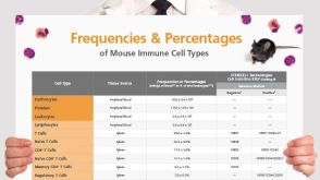 挂图Frequencies and Percentages of Mouse Immune Cell Types List of the frequencies of over 25 immune cell types in C57BL/6 mice
 沪公网安备31010102008431号