Griffin DO et al. (JAN 2011)
The Journal of experimental medicine 208 1 67--80
Human B1 cells in umbilical cord and adult peripheral blood express the novel phenotype CD20+ CD27+ CD43+ CD70-.
B1 cells differ in many ways from conventional B cells,most prominently in the production of natural immunoglobulin,which is vitally important for protection against pathogens. B1 cells have also been implicated in the pathogenesis of autoimmune dyscrasias and malignant diseases. It has been impossible to accurately study B1 cells during health and illness because the nature of human B1 cells has not been successfully defined. This has produced controversy regarding the existence of human B1 cells. Here,we determined the phenotype of human B1 cells by testing sort-purified B cell fractions for three fundamental B1 cell functions based on mouse studies: spontaneous IgM secretion,efficient T cell stimulation,and tonic intracellular signaling. We found that a small population of CD20(+)CD27(+)CD43(+) cells present in both umbilical cord and adult peripheral blood fulfilled these criteria and expressed a skewed B cell receptor repertoire. These B cells express little or no surface CD69 and CD70,both of which are markedly up-regulated after activation of CD20(+)CD27(-)CD43(-) (naive) and CD20(+)CD27(+)CD43(-) (memory) B cells. This work identifies human B1 cells as CD20(+)CD27(+)CD43(+)CD70(-). We determined that the proportion of B1 cells declines with age,which may contribute to disease susceptibility. Identification of human B1 cells provides a foundation for future studies on the nature and role of these cells in human disease.
View Publication
产品号#:
18054
18054RF
19155
19155RF
产品名:
C.-W. J. Lio et al. (apr 2019)
Science immunology 4 34
TET enzymes augment activation-induced deaminase (AID) expression via 5-hydroxymethylcytosine modifications at the Aicda superenhancer.
TET enzymes are dioxygenases that promote DNA demethylation by oxidizing the methyl group of 5-methylcytosine to 5-hydroxymethylcytosine (5hmC). Here,we report a close correspondence between 5hmC-marked regions,chromatin accessibility and enhancer activity in B cells,and a strong enrichment for consensus binding motifs for basic region-leucine zipper (bZIP) transcription factors at TET-responsive genomic regions. Functionally,Tet2 and Tet3 regulate class switch recombination (CSR) in murine B cells by enhancing expression of Aicda,which encodes the activation-induced cytidine deaminase (AID) enzyme essential for CSR. TET enzymes deposit 5hmC,facilitate DNA demethylation,and maintain chromatin accessibility at two TET-responsive enhancer elements,TetE1 and TetE2,located within a superenhancer in the Aicda locus. Our data identify the bZIP transcription factor,ATF-like (BATF) as a key transcription factor involved in TET-dependent Aicda expression. 5hmC is not deposited at TetE1 in activated Batf-deficient B cells,indicating that BATF facilitates TET recruitment to this Aicda enhancer. Our study emphasizes the importance of TET enzymes for bolstering AID expression and highlights 5hmC as an epigenetic mark that captures enhancer dynamics during cell activation.
View Publication
产品号#:
19854
19854RF
产品名:
EasySep™小鼠B细胞分选试剂盒
RoboSep™ 小鼠B细胞分选试剂盒
J. U. Hermansen et al. (dec 2018)
Scientific reports 8 1 17651
Cryopreservation of primary B cells minimally influences their signaling responses.
Phospho flow is a powerful approach to detect cell signaling aberrations,identify biomarkers and assess pharmacodynamics,and can be performed using cryopreserved samples. The effects of cryopreservation on signaling responses and the reproducibility of phospho flow measurements are however unknown in many cell systems. Here,B lymphocytes were isolated from healthy donors and patients with the B cell malignancy chronic lymphocytic leukemia and analyzed by phospho flow using phospho-specific antibodies targeting 20 different protein epitopes. Cells were analyzed both at basal conditions and after activation of cluster of differentiation 40 (CD40) or the B cell receptor. Pharmacodynamics of the novel pathway inhibitor ibrutinib was also assessed. At all conditions,fresh cells were compared to cryopreserved cells. Minimal variation between fresh and frozen samples was detected. Reproducibility was tested by running samples from the same donors in different experiments. The results demonstrate reproducibility across different phospho flow runs and support the use of cryopreserved samples in future phospho flow studies of B lymphocytes.
View Publication
产品号#:
15024
15064
产品名:
RosetteSep™人B细胞富集抗体混合物
RosetteSep™人B细胞富集抗体混合物
de Valle E et al. (APR 2016)
The Journal of Experimental Medicine 213 4 621--41
NFκB1 is essential to prevent the development of multiorgan autoimmunity by limiting IL-6 production in follicular B cells.
We examined the role of NFκB1 in the homeostasis and function of peripheral follicular (Fo) B cells. Aging mice lacking NFκB1 (Nfκb1(-/-)) develop lymphoproliferative and multiorgan autoimmune disease attributed in large part to the deregulated activity ofNfκb1(-/-)Fo B cells that produce excessive levels of the proinflammatory cytokine interleukin 6 (IL-6). Despite enhanced germinal center (GC) B cell differentiation,the formation of GC structures was severely disrupted in theNfκb1(-/-)mice. Bone marrow chimeric mice revealed that the Fo B cell-intrinsic loss of NFκB1 led to the spontaneous generation of GC B cells. This was primarily the result of an increase in IL-6 levels,which promotes the differentiation of Fo helper CD4(+)T cells and acts in an autocrine manner to reduce antigen receptor and toll-like receptor activation thresholds in a population of proliferating IgM(+)Nfκb1(-/-)Fo B cells. We demonstrate that p50-NFκB1 repressesIl-6transcription in Fo B cells,with the loss of NFκB1 also resulting in the uncontrolled RELA-driven transcription ofIl-6.Collectively,our findings identify a previously unrecognized role for NFκB1 in preventing multiorgan autoimmunity through its negative regulation ofIl-6gene expression in Fo B cells.
View Publication
产品号#:
19854
19854RF
产品名:
EasySep™小鼠B细胞分选试剂盒
RoboSep™ 小鼠B细胞分选试剂盒
Flach A-C et al. (MAR 2016)
Proceedings of the National Academy of Sciences of the United States of America 113 12 3323--8
Autoantibody-boosted T-cell reactivation in the target organ triggers manifestation of autoimmune CNS disease.
Multiple sclerosis (MS) is caused by T cells that are reactive for brain antigens. In experimental autoimmune encephalomyelitis,the animal model for MS,myelin-reactive T cells initiate the autoimmune process when entering the nervous tissue and become reactivated upon local encounter of their cognate CNS antigen. Thereby,the strength of the T-cellular reactivation process within the CNS tissue is crucial for the manifestation and the severity of the clinical disease. Recently,B cells were found to participate in the pathogenesis of CNS autoimmunity,with several diverse underlying mechanisms being under discussion. We here report that B cells play an important role in promoting the initiation process of CNS autoimmunity. Myelin-specific antibodies produced by autoreactive B cells after activation in the periphery diffused into the CNS together with the first invading pathogenic T cells. The antibodies accumulated in resident antigen-presenting phagocytes and significantly enhanced the activation of the incoming effector T cells. The ensuing strong blood-brain barrier disruption and immune cell recruitment resulted in rapid manifestation of clinical disease. Therefore,myelin oligodendrocyte glycoprotein (MOG)-specific autoantibodies can initiate disease bouts by cooperating with the autoreactive T cells in helping them to recognize their autoantigen and become efficiently reactivated within the immune-deprived nervous tissue.
View Publication
Rovituso DM et al. ( 2016)
Scientific reports 6 29847
CEACAM1 mediates B cell aggregation in central nervous system autoimmunity.
B cell aggregates in the central nervous system (CNS) have been associated with rapid disease progression in patients with multiple sclerosis (MS). Here we demonstrate a key role of carcinoembryogenic antigen-related cell adhesion molecule1 (CEACAM1) in B cell aggregate formation in MS patients and a B cell-dependent mouse model of MS. CEACAM1 expression was increased on peripheral blood B cells and CEACAM1(+) B cells were present in brain infiltrates of MS patients. Administration of the anti-CEACAM1 antibody T84.1 was efficient in blocking aggregation of B cells derived from MS patients. Along these lines,application of the monoclonal anti-CEACAM1 antibody mCC1 was able to inhibit CNS B cell aggregate formation and significantly attenuated established MS-like disease in mice in the absence of any adverse effects. CEACAM1 was co-expressed with the regulator molecule T cell immunoglobulin and mucin domain -3 (TIM-3) on B cells,a novel molecule that has recently been described to induce anergy in T cells. Interestingly,elevated coexpression on B cells coincided with an autoreactive T helper cell phenotype in MS patients. Overall,these data identify CEACAM1 as a clinically highly interesting target in MS pathogenesis and open new therapeutic avenues for the treatment of the disease.
View Publication
产品号#:
15024
15064
产品名:
RosetteSep™人B细胞富集抗体混合物
RosetteSep™人B细胞富集抗体混合物
Chorny A et al. (SEP 2016)
The Journal of experimental medicine
The soluble pattern recognition receptor PTX3 links humoral innate and adaptive immune responses by helping marginal zone B cells.
Pentraxin 3 (PTX3) is a fluid-phase pattern recognition receptor of the humoral innate immune system with ancestral antibody-like properties but unknown antibody-inducing function. In this study,we found binding of PTX3 to splenic marginal zone (MZ) B cells,an innate-like subset of antibody-producing lymphocytes strategically positioned at the interface between the circulation and the adaptive immune system. PTX3 was released by a subset of neutrophils that surrounded the splenic MZ and expressed an immune activation-related gene signature distinct from that of circulating neutrophils. Binding of PTX3 promoted homeostatic production of IgM and class-switched IgG antibodies to microbial capsular polysaccharides,which decreased in PTX3-deficient mice and humans. In addition,PTX3 increased IgM and IgG production after infection with blood-borne encapsulated bacteria or immunization with bacterial carbohydrates. This immunogenic effect stemmed from the activation of MZ B cells through a neutrophil-regulated pathway that elicited class switching and plasmablast expansion via a combination of T cell-independent and T cell-dependent signals. Thus,PTX3 may bridge the humoral arms of the innate and adaptive immune systems by serving as an endogenous adjuvant for MZ B cells. This property could be harnessed to develop more effective vaccines against encapsulated pathogens.
View Publication
产品号#:
19754
19754RF
产品名:
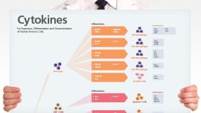
挂图
Human Immune Cytokines
Infographic of key cytokines for expansion, differentiation and characterization of major immune cell types
Gilbert AE et al. (JAN 2011)
PloS one 6 4 e19330
Monitoring the systemic human memory B cell compartment of melanoma patients for anti-tumor IgG antibodies.
Melanoma,a potentially lethal skin cancer,is widely thought to be immunogenic in nature. While there has been much focus on T cell-mediated immune responses,limited knowledge exists on the role of mature B cells. We describe an approach,including a cell-based ELISA,to evaluate mature IgG antibody responses to melanoma from human peripheral blood B cells. We observed a significant increase in antibody responses from melanoma patients (n = 10) to primary and metastatic melanoma cells compared to healthy volunteers (n = 10) (Ptextless0.0001). Interestingly,we detected a significant reduction in antibody responses to melanoma with advancing disease stage in our patient cohort (n = 21) (Ptextless0.0001). Overall,28% of melanoma patient-derived B cell cultures (n = 1,800) compared to 2% of cultures from healthy controls (n = 600) produced antibodies that recognized melanoma cells. Lastly,a patient-derived melanoma-specific monoclonal antibody was selected for further study. This antibody effectively killed melanoma cells in vitro via antibody-mediated cellular cytotoxicity. These data demonstrate the presence of a mature systemic B cell response in melanoma patients,which is reduced with disease progression,adding to previous reports of tumor-reactive antibodies in patient sera,and suggesting the merit of future work to elucidate the clinical relevance of activating humoral immune responses to cancer.
View Publication
产品号#:
15024
15064
产品名:
RosetteSep™人B细胞富集抗体混合物
RosetteSep™人B细胞富集抗体混合物
Nova-Lamperti E et al. (JAN 2016)
Scientific Reports 6 20044
IL-10-produced by human transitional B-cells down-regulates CD86 expression on B-cells leading to inhibition of CD4+T-cell responses.
A novel subset of human regulatory B-cells has recently been described. They arise from within the transitional B-cell subpopulation and are characterised by the production of IL-10. They appear to be of significant importance in regulating T-cell immunity in vivo. Despite this important function,the molecular mechanisms by which they control T-cell activation are incompletely defined. Here we show that transitional B-cells produced more IL-10 and expressed higher levels of IL-10 receptor after CD40 engagement compared to other B-cell subsets. Furthermore,under this stimulatory condition,CD86 expressed by transitional B-cells was down regulated and T-cell proliferation was reduced. We provide evidence to demonstrate that the down-regulation of CD86 expression by transitional B-cells was due to the autocrine effect of IL-10,which in turn leads to decreased T-cell proliferation and TNF-α production. This analysis was further extended to peripheral B-cells in kidney transplant recipients. We observed that B-cells from patients tolerant to the graft maintained higher IL-10 production after CD40 ligation,which correlates with lower CD86 expression compared to patients with chronic rejection. Hence,the results obtained in this study shed light on a new alternative mechanism by which transitional B-cells inhibit T-cell proliferation and cytokine production.
View Publication
产品号#:
15022
15062
15024
15064
产品名:
RosetteSep™人CD4+ T细胞富集抗体混合物
RosetteSep™人CD4+ T细胞富集抗体混合物
RosetteSep™人B细胞富集抗体混合物
RosetteSep™人B细胞富集抗体混合物
Valsecchi R et al. (APR 2016)
Blood 127 16 1987--97
HIF-1α regulates the interaction of chronic lymphocytic leukemia cells with the tumor microenvironment.
Hypoxia-inducible transcription factors (HIFs) regulate a wide array of adaptive responses to hypoxia and are often activated in solid tumors and hematologic malignancies due to intratumoral hypoxia and emerging new layers of regulation. We found that in chronic lymphocytic leukemia (CLL),HIF-1α is a novel regulator of the interaction of CLL cells with protective leukemia microenvironments and,in turn,is regulated by this interaction in a positive feedback loop that promotes leukemia survival and propagation. Through unbiased microarray analysis,we found that in CLL cells,HIF-1α regulates the expression of important chemokine receptors and cell adhesion molecules that control the interaction of leukemic cells with bone marrow and spleen microenvironments. Inactivation of HIF-1α impairs chemotaxis and cell adhesion to stroma,reduces bone marrow and spleen colonization in xenograft and allograft CLL mouse models,and prolongs survival in mice. Of interest,we found that in CLL cells,HIF-1α is transcriptionally regulated after coculture with stromal cells. Furthermore,HIF-1α messenger RNA levels vary significantly within CLL patients and correlate with the expression of HIF-1α target genes,including CXCR4,thus further emphasizing the relevance of HIF-1α expression to CLL pathogenesis.
View Publication

 EasySep™小鼠TIL(CD45)正选试剂盒


 挂图Human Immune Cytokines Infographic of key cytokines for expansion, differentiation and characterization of major immune cell types

 沪公网安备31010102008431号