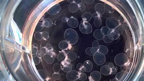
Nanofiber-microwell cell culture system for spatially patterned differentiation of pluripotent stem cells in 3D
The intricate interplay between biochemical and physical cues dictates pluripotent stem cell (PSC) differentiation to form various tissues. While biochemical modulation has been extensively studied,the role of biophysical microenvironments in early lineage commitment remains elusive. Here,we introduce a novel 3D cell culture system combining electrospun nanofibers with microfabricated polydimethylsiloxane (PDMS) patterns. This system enables the controlled formation of semispherical human induced pluripotent stem cell (hiPSC) colonies,facilitating the investigation of local mechanical stem cell niches on mechano-responsive signaling and lineage specification. Our system unveiled spatially organized RhoA activity coupled with actin-myosin cable formation,suggesting mechano-dependent hiPSC behaviors. Nodal network analysis of RNA-seq data revealed RhoA downstream regulation of YAP signaling,DNA histone modifications,and patterned germ layer specification. Notably,altering colony morphology through controlled PDMS microwell shaping effectively modulated the spatial distribution of mechano-sensitive mediators and subsequent differentiation. This study provides a cell culture platform to decipher the role of biophysical cues in early embryogenesis,offering valuable insights for material design in tissue engineering and regenerative medicine applications. Graphical abstractImage 1
View Publication
产品号#:
85850
85857
产品名:
mTeSR™1
mTeSR™1
(Oct 2024)
BMC Psychiatry 24 1
Patient iPSC-derived neural progenitor cells display aberrant cell cycle control, p53, and DNA damage response protein expression in schizophrenia
BackgroundSchizophrenia (SCZ) is a severe psychiatric disorder associated with alterations in early brain development. Details of underlying pathomechanisms remain unclear,despite genome and transcriptome studies providing evidence for aberrant cellular phenotypes and pathway deregulation in developing neuronal cells. However,mechanistic insight at the protein level is limited.MethodsHere,we investigate SCZ-specific protein expression signatures of neuronal progenitor cells (NPC) derived from patient iPSC in comparison to healthy controls using high-throughput Western Blotting (DigiWest) in a targeted proteomics approach.ResultsSCZ neural progenitors displayed altered expression and phosphorylation patterns related to Wnt and MAPK signaling,protein synthesis,cell cycle regulation and DNA damage response. Consistent with impaired cell cycle control,SCZ NPCs also showed accumulation in the G2/M cell phase and reduced differentiation capacity. Furthermore,we correlated these findings with elevated p53 expression and phosphorylation levels in SCZ patient-derived cells,indicating a potential implication of p53 in hampering cell cycle progression and efficient neurodevelopment in SCZ.ConclusionsThrough targeted proteomics we demonstrate that SCZ NPC display coherent mechanistic alterations in regulation of DNA damage response,cell cycle control and p53 expression. These findings highlight the suitability of iPSC-based approaches for modeling psychiatric disorders and contribute to a better understanding of the disease mechanisms underlying SCZ,particularly during early development.Supplementary InformationThe online version contains supplementary material available at 10.1186/s12888-024-06127-x.
View Publication
产品号#:
05833
08581
08582
100-0276
100-1130
产品名:
STEMdiff™神经前体细胞培养基
STEMdiff™SMADi神经诱导试剂盒
STEMdiff™SMADi神经诱导试剂盒,2套
mTeSR™ Plus
mTeSR™ Plus
(Apr 2025)
Cells 14 7
Induced Pluripotent Stem Cell-Derived Exosomes Promote Peripheral Nerve Regeneration in a Rat Sciatic Nerve Crush Injury Model: A Safety and Efficacy Study
Peripheral nerve injury (PNI) remains a significant clinical challenge,often leading to long-term functional impairment. Despite advances in therapies,current repair strategies offer unsatisfactory clinical outcomes. Exosomes derived from induced pluripotent stem cells (iPSC-Exos) have emerged as a promising therapeutic approach in regenerative medicine. This study assesses the efficacy and safety of iPSC-Exos in a rat model of sciatic nerve crush injury. Briefly,iPSCs were generated from peripheral blood mononuclear cells (PBMCs) of healthy donors using Sendai virus vectors and validated for pluripotency. iPSC-Exos were characterized and injected at the injury site. Functional recovery was assessed through gait analysis,grip strength,and pain response. Histological and molecular analyses were used to examine axonal regeneration,myelination,Schwann cell (SC) activation,angiogenesis,and changes in gene expression. iPSC-Exos were efficiently internalized by SC,promoting their proliferation. No adverse effects were observed between groups on body weight,organ histology,or hematological parameters. iPSC-Exos injection significantly enhanced nerve regeneration,muscle preservation,and vascularization,with RNA sequencing revealing activation of PI3K-AKT and focal adhesion pathways. These findings support iPSC-Exos as a safe and effective non-cell-based therapy for PNIs,highlighting their potential for clinical applications in regenerative medicine.
View Publication

 EasySep™小鼠TIL(CD45)正选试剂盒


 专家访谈Dr. Nika Shakiba From Mentee to Mentor: How to Help Trainees Thrive,with Dr. Nika Shakiba from UBC


 沪公网安备31010102008431号