C. Nguyen et al. (Oct 2025)
Nature Immunology 26 11
Transcriptional and epigenetic targets of MEF2C in human microglia contribute to cellular functions related to autism risk and age-related disease
MEF2C encodes a transcription factor that is critical in nervous system development. Here,to examine disease-associated functions of MEF2C in human microglia,we profiled microglia differentiated from isogenic MEF2C-haploinsufficient and MEF2C-knockout induced pluripotent stem cell lines. Complementary transcriptomic and functional analyses revealed that loss of MEF2C led to a hyperinflammatory phenotype with broad phagocytic impairment,lipid accumulation,lysosomal dysfunction and elevated basal inflammatory cytokine secretion. Genome-wide profiling of MEF2C-bound sites coupled with the active regulatory landscape enabled inference of its transcriptional functions and potential mechanisms for MEF2C-associated cellular functions. Transcriptomic and epigenetic approaches identified substantial overlap with idiopathic autism datasets,suggesting a broader role of human microglial MEF2C dysregulation in idiopathic autism. In a mouse xenotransplantation model,loss of MEF2C led to morphological,lysosomal and lipid abnormalities in human microglia in vivo. Together,these studies reveal mechanisms by which reduced microglial MEF2C could contribute to the development of neurological diseases. Coufal and colleagues generated microglia from human iPS cells to examine mechanistic roles of the transcription factor MEF2C and how these roles might relate to the autism phenotype seen following the loss of MEF2C in human microglia.
View Publication
产品号#:
100-0483
100-0484
产品名:
Hausser Scientificᵀᴹ 明线血球计数板
ReLeSR™
R. U. W. Friis et al. (Oct 2025)
Cancer Immunology,Immunotherapy : CII 74 11
Development of antigen multimers for detection and evaluation of CAR T cells
Chimeric antigen receptor (CAR) T cell therapy has transformed the treatment landscape of hematologic cancers by engineering T cells to specifically target and destroy cancer cells. Monitoring CAR T cell activity and function is essential for optimizing therapeutic outcomes,but existing tools for CAR detection are often limited in specificity and functional assessment capability. Methods: We developed dextran multimers by conjugating multiple CAR-specific antigens to a dextran backbone. The multimers were compared to previously reported antigen tetramers for their ability to stain and detect CAR T cells. Because these multimers incorporate the CAR target antigen,they uniquely enable assessment of CAR T cell functionality. We tested the staining and functional properties of the multimers across a range of CAR constructs with different affinities,using flow cytometry and microscopy. Results: The dextran multimers demonstrated high specificity and sensitivity in staining CAR T cells,with adjustable antigen density to optimize binding. Dextran multimers also enabled effective clustering and subsequent activation of CARs,showing their utility as both a staining and functional assessment tool. The multimers revealed that CARs with different affinities and clustering tendencies displayed varied binding and activation in response to different antigen densities. Conclusions: Dextran multimers offer a dual advantage as versatile reagents for both staining and functional analysis of CAR T cells. Their capacity to engage CARs with the specific antigen provides a valuable platform for evaluating CAR functionality,informing CAR design improvements,and enhancing therapeutic precision.
View Publication
产品号#:
100-0695
17951
17951RF
产品名:
EasySep™人T细胞分选试剂盒
EasySep™人T细胞分选试剂盒
RoboSep™ 人T细胞分选试剂盒
Y. Fan et al. (Oct 2025)
Cell & Bioscience 15 5819
Breaking the link between morphology and potency for mESCs
In stem cell biology,a long-held structure–function relationship is the domed colony morphology and naïve pluripotency for mouse or human pluripotent stem cells. This link has provided a convenient way to recognize bona fide naïve pluripotent cells during derivation,passaging and characterization. However,the molecular basis of this link remains poorly understood. Results: We show that a loss of domed morphology may not impact the overall genetic architecture of naïve pluripotency in mouse embryonic stem cells (mESCs). We first generated stable mESC lines by knocking out Myh9 that encodes non-muscle myosin heavy chain IIA,resulting in colonies deprived of the typical domed morphology,but competent to differentiate into the three germ layers and chimeric mice. Modulating cell morphologies with inhibitors against kinases known to regulate myosin pathway also phenocopy the knockout in wild type mESCs. Conclusions: These results provide evidence that the domed morphology and potency can be uncoupled and suggest that domed structure is not a pre-requisite for acquiring and maintaining naïve pluripotency.
View Publication
产品号#:
85850
85857
产品名:
mTeSR™1
mTeSR™1
P. Dai et al. (Oct 2025)
Clinical & Translational Immunology 14 10
A semi‐automated ASC speck assay to evaluate pyrin inflammasome activation
Objective: To develop a rapid functional assay to validate variants of uncertain significance (VUS) in the MEFV gene. Methods: Overactivity of the pyrin inflammasome pathway and ASC speck oligomerisation in response to stimulation with low concentrations of Clostridium difficile toxin A was directly visualised by immunofluorescence microscopy. A semi‐automated algorithm was developed to count cells and ASC specks. Results: The semi‐automated ASC speck assay is able to discriminate between healthy controls and patients with familial Mediterranean fever (FMF) and pyrin inflammasome overactivity with high sensitivity. It is also able to discriminate pyrin inflammasome overactivity from other autoinflammatory disease controls with high specificity. Conclusion: The semi‐automated ASC speck assay may be a useful test to functionally validate VUS in the MEFV gene and screen for pyrin inflammasome overactivity. A semi‐automated ASC speck assay using machine learning is able to discriminate between healthy controls and patients with familial Mediterranean fever (FMF) with high sensitivity. It is also able to discriminate FMF from other autoinflammatory diseases with high specificity.
View Publication
产品号#:
100-0694
17858
17858RF
产品名:
EasySep™人CD14正选试剂盒II
EasySep™人CD14正选试剂盒II
RoboSep™ 人CD14正选试剂盒II
A. Demchenko et al. (Oct 2025)
PLOS Computational Biology 21 10
A semi-automated algorithm for image analysis of respiratory organoids
Respiratory organoids have emerged as a powerful in vitro model for studying respiratory diseases and drug discovery. However,the high-throughput analysis of organoid images remains a challenge due to the lack of automated and accurate segmentation tools. This study presents a semi-automatic algorithm for image analysis of respiratory organoids (nasal and lung organoids),employing the U-Net architecture and CellProfiler for organoids segmentation. The algorithm processes bright-field images acquired through z-stack fusion and stitching. The model demonstrated a high level of accuracy,as evidenced by an intersection-over-union metric (IoU) of 0.8856,F1-score = 0.937 and an accuracy of 0.9953. Applied to forskolin-induced swelling assays of lung organoids,the algorithm successfully quantified functional differences in Cystic Fibrosis Transmembrane conductance Regulator (CFTR)-channel activity between healthy donor and cystic fibrosis patient-derived organoids,without fluorescent dyes. Additionally,an open-source dataset of 827 annotated respiratory organoid images was provided to facilitate further research. Our results demonstrate the potential of deep learning to enhance the efficiency and accuracy of high-throughput respiratory organoid analysis for future therapeutic screening applications. Author summaryIn this study,we developed a semi-automated tool to analyze images of respiratory organoids—3D cell structures that mimic the human respiratory system. These organoids are vital for studying diseases like cystic fibrosis and testing potential drugs,but manually analyzing their images is time-consuming and prone to errors. Our tool uses artificial intelligence (AI) to quickly and accurately measure organoid size and shape from bright-field microscope images,eliminating the need for fluorescent dyes that can harm cells. We trained our AI model on a publicly shared dataset of 827 annotated organoid images,achieving high accuracy in detecting and quantifying organoids. When applied to cystic fibrosis research,the tool successfully measured differences in organoid swelling (forskolin-induced swelling - a key test for drug response) between healthy and patient-derived samples. By making our dataset and method openly available,we hope to support further research into respiratory diseases. Our work bridges the gap between complex lab techniques and practical applications,offering a faster,more reliable way to study human health and disease.
View Publication
产品号#:
05040
产品名:
PneumaCult™-Ex Plus 培养基
H. Kurniawan et al. (Oct 2025)
Journal of Neuroinflammation 22 23
The Parkinson’s disease-associated LRRK2-G2019S variant restricts serine metabolism, leading to microglial inflammation and dopaminergic neuron degeneration
A growing body of evidence implicates inflammation as a key hallmark in the pathophysiology of Parkinson’s disease (PD),with microglia playing a central role in mediating neuroinflammatory signaling in the brain. However,the molecular mechanisms linking microglial activation to dopaminergic neuron degeneration remain poorly understood. In this study,we investigated the contribution of the PD-associated LRRK2-G2019S mutation to microglial neurotoxicity using patient-derived induced pluripotent stem cell (iPSC) models. We found that LRRK2-G2019S mutant microglia exhibited elevated activation markers,enhanced phagocytic capacity,and increased secretion of pro-inflammatory cytokines such as TNF-α. These changes were associated with metabolic dysregulation,including upregulated glycolysis and impaired serine biosynthesis. In 3D midbrain organoids,these overactivated microglia resulted in dopaminergic neuron degeneration. Notably,treating LRRK2-G2019S microglia with oxamic acid,a glycolysis inhibitor,attenuated microglial inflammation and reduced neuronal loss. Our findings underscore the link between metabolic targeting in microglia and dopaminergic neuronal loss in LRRK2-G2019S mutation,and highlight a potential strategy that warrants further preclinical evaluation.
View Publication
产品号#:
34811
34815
34821
34825
34850
34860
产品名:
AggreWell™ 800 24孔板,1个
AggreWell™ 800 24孔板,5个
AggreWell™ 800 6孔板,1个
AggreWell™ 800 6孔板,5个
AggreWell™ 800 24孔板启动套装
AggreWell™ 800 6孔板启动套装
D. Zheng et al. (Oct 2025)
Stem Cell Research & Therapy 16
Dynamic molecular and cellular characteristics of VSX2-positive retinal progenitor cells in human retinal organoids
The lack of understanding of the molecular and cellular characteristics of human retinal progenitor cells (RPCs) has hindered their application in cell therapy for retinal degenerative diseases. This study aims to employ retinal organoids (ROs) derived from a VSX2-enhanced green fluorescent protein (eGFP) reporter human induced pluripotent stem cell (hiPSC) line for positive selection of human RPCs,investigate their features,and facilitate their applications. Methods: hiPSCs were differentiated into three-dimensional ROs following established protocols. The fidelity of the VSX2-eGFP reporter was confirmed through immunostaining. Fluorescence-activated cell sorting was employed to select VSX2-eGFP-positive (+) cells at distinct developmental stages,followed by bulk RNA sequencing (RNA-seq) analysis to assess their transcriptome profile. Immunostaining and flow cytometry were utilized to validate the identity of VSX2-eGFP+ cells and potential cluster of differentiation (CD) biomarkers for identifying human RPCs. Results: hiPSCs were successfully differentiated into ROs containing abundant RPCs. The spatiotemporal activity of the VSX2-eGFP reporter recapitulated the dynamic expression of endogenous VSX2 protein. Compared to VSX2-eGFP-negative (-) cells,VSX2-eGFP+ cells mainly exhibited characteristics of RPCs at early stages of retinal development and of bipolar cells at late stages. RNA-seq analysis revealed transcriptional heterogeneity within VSX2-eGFP+ cells across four distinct developmental stages. Moreover,the dynamic expression of 394 known CD biomarkers in VSX2-eGFP+ cells at distinct developmental stages was analyzed herein for the first time. One CD biomarker,TNFRSF1B,which has never been reported to be expressed in RPCs,was found to be highly expressed in RPCs at the early stages and might serve as a candidate CD biomarker for sorting RPCs. Conclusions: This study provides valuable insights into the molecular and cellular characteristics of human RPCs,especially their expression profiles of CD biomarkers,laying a foundation for research on retinal development and the clinical translation of hiPSC-derived RPCs.
View Publication

 EasySep™小鼠TIL(CD45)正选试剂盒


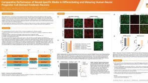 科学海报Comparative Performance of Neural-Specific Media in Differentiating and Maturing Human Neural Progenitor Cell-Derived Forebrain Neurons
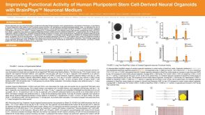 科学海报Improving Functional Activity of Human Pluripotent Stem Cell-Derived Neural Organoids with BrainPhys Neuronal Medium
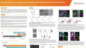 科学海报Optimized Workflows for Modelling Human Pluripotent Stem Cell-Derived Neuron and Glial Interactions

 沪公网安备31010102008431号