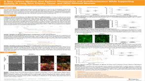
Simple and versatile synthetic polydopamine-based surface supports reprogramming of human somatic cells and long-term self-renewal of human pluripotent stem cells under defined conditions
Human pluripotent stem cells (hPSCs) possess great value in the aspect of cellular therapies due to its self-renewal and potential to differentiate into all somatic cell types. A few defined synthetic surfaces such as polymers and adhesive biological materials conjugated substrata were established for the self-renewal of hPSCs. However,none of them was effective in the generation of human induced pluripotent stem cells (hiPSCs) and long-term maintenance of multiple hPSCs,and most of them required complicated manufacturing processes. Polydopamine has good biocompatibility,is able to form a stable film on nearly all solid substrates surface,and can immobilize adhesive biomolecules. In this manuscript,a polydopamine-mediated surface was developed,which not only supported the reprogramming of human somatic cells into hiPSCs under defined conditions,but also sustained the growth of hiPSCs on diverse substrates. Moreover,the proliferation and pluripotency of hPSCs cultured on the surface were comparable to Matrigel for more than 20 passages. Besides,hPSCs were able to differentiate to cardiomyocytes and neural cells on the surface. This polydopamine-based synthetic surface represents a chemically-defined surface extensively applicable both for fundamental research and cell therapies of hPSCs.
View Publication
Katori S et al. (JUL 2009)
The Journal of neuroscience : the official journal of the Society for Neuroscience 29 29 9137--47
Protocadherin-alpha family is required for serotonergic projections to appropriately innervate target brain areas.
Serotonergic axons from the raphe nuclei in the brainstem project to every region of the brain,where they make connections through their extensive terminal arborizations. This serotonergic innervation contributes to various normal behaviors and psychiatric disorders. The protocadherin-alpha (Pcdha) family of clustered protocadherins consists of 14 cadherin-related molecules generated from a single gene cluster. We found that the Pcdhas were strongly expressed in the serotonergic neurons. To elucidate their roles,we examined serotonergic fibers in a mouse mutant (Pcdha(Delta CR/Delta CR)) lacking the Pcdha cytoplasmic region-encoding exons,which are common to the gene cluster. In the first week after birth,the distribution pattern of serotonergic fibers in Pcdha(Delta CR/Delta CR) mice was similar to wild-type,but by 3 weeks of age,when the serotonergic axonal termini complete their arborizations,the distribution of the projections was abnormal. In some target regions,notably the globus pallidus and substantia nigra,the normally even distribution of serotonin axonal terminals was,in the mutants,dense at the periphery of each region,but sparse in the center. In the stratum lacunosum-molecular of the hippocampus,the mutants showed denser serotonergic innervation than in wild-type,and in the dentate gyrus of the hippocampus and the caudate-putamen,the innervation was sparser. Together,the abnormalities suggested that Pcdha proteins are important in the late-stage maturation of serotonergic projections. Further examination of alternatively spliced exons encoding the cytoplasmic tail showed that the A-type (but not the B-type) cytoplasmic tail was essential for the normal development of serotonergic projections.
View Publication
产品号#:
03800
03801
03802
03803
03804
03805
03806
产品名:
ClonaCell™-HY杂交瘤试剂盒
ClonaCell™-HY培养基A
ClonaCell™-HY 培养基 B
ClonaCell™-HY 培养基 C
ClonaCell™-HY 培养基 D
ClonaCell™-HY 培养基 E
ClonaCell™-HY PEG
Jackson TC et al. (FEB 2018)
Experimental Neurology 300 232--246
BrainPhys increases neurofilament levels in CNS cultures, and facilitates investigation of axonal damage after a mechanical stretch-injury in vitro
Neurobasal®/B27 is a gold standard culture media used to study primary neurons in vitro. An alternative media (BrainPhys®/SM1) was recently developed which robustly enhances neuronal activity vs. Neurobasal® or DMEM. To the best of our knowledge BrainPhys® has not been explored in the setting of neuronal injury. Here we characterized the utility of BrainPhys® in a model of in vitro mechanical-stretch injury. METHODS/RESULTSPrimary rat cortical neurons were maintained in classic Neurobasal®,or sequentially maintained in Neurocult® followed by BrainPhys® (hereafter simply referred to as BrainPhys® maintained neurons?). The levels of axonal markers and proteins involved in neurotransmission were compared on day in vitro 10 (DIV10). BrainPhys® maintained neurons had higher levels of GluN2B,GluR1,Neurofilament light/heavy chain (NF-L & NF-H),and protein phosphatase 2 A (PP2A) vs. neurons in Neurobasal®. Mechanical stretch-injury (50ms/54% biaxial stretch) to BrainPhys® maintained neurons modestly (albeit significantly) increased 24h lactate dehydrogenase (LDH) levels but markedly decreased axonal NF-L levels post-injury vs. uninjured controls or neurons given a milder 38% stretch-injury. Furthermore,two 54% stretch-injuries (in tandem) exacerbated 24h LDH release,increased α-spectrin breakdown products (SBDPs),and decreased Tau levels. Also,BrainPhys® maintained cultures had decreased markers of cell damage 24h after a single 54% stretch-injury vs. neurons in Neurobasal®. Finally,we tested the hypothesis that lentivirus mediated overexpression of the pro-death protein RBM5 exacerbates neuronal and/or axonal injury in primary CNS cultures. RBM5 overexpression vs. empty-vector controls increased 24h LDH release,and SBDP levels,after a single 54% stretch-injury but did not affect NF-L levels or Tau. CONCLUSIONBrainPhys® is a promising new reagent which facilities the investigation of molecular targets involved in axonal and/or neuronal injury in vitro.
View Publication
产品号#:
05790
05792
05793
05794
05795
产品名:
BrainPhys™神经元培养基
BrainPhys™神经元培养基和SM1试剂盒
BrainPhys™ 神经元培养基N2-A和SM1试剂盒
BrainPhys™原代神经元试剂盒
BrainPhys™ hPSC 神经元试剂盒
Jessick VJ et al. ( 2013)
International journal of physiology,pathophysiology and pharmacology 5 4 216--27
Investigating the role of the actin regulating complex ARP2/3 in rapid ischemic tolerance induced neuro-protection.
Neuronal morphology is highly sensitive to ischemia,although some re-organization may promote neuroprotection. In this study we investigate the role of actin regulating proteins (ARP2,ARP3 and WAVE-1) and their role in rapid ischemic tolerance. Using an established in vitro model of rapid ischemic tolerance,we show that WAVE-1 protein levels are stabilized following brief tolerance inducing ischemia (preconditioning). The stabilization appears to be due to a reduction in the ubiquitination of WAVE-1. Levels of ARP2,ARP3 and N-WASP were not affected by ischemic preconditioning. Immunocytochemical studies show a relocalization of ARP2 and ARP3 proteins in neurons following preconditioning ischemia,as well as a re-organization of actin. Blocking the protein kinase CK2 using emodin blocks ischemic tolerance,and our data suggests CK2 binds to WAVE-1 in neurons. We observe an increase in binding of the ARP2 subunit with WAVE-1. The neuroprotection observed following preconditioning is inhibited when cells are transduced with an N-WASP CA domain that blocks the activation of ARP2/3. Together these data show that ischemia affects actin regulating enzymes,and that the ARP2/3 pathway plays a role in rapid ischemic tolerance induced neuroprotection.
View Publication
产品号#:
05711
100-1281
产品名:
NeuroCult™ SM1 神经添加物
NeuroCult™ SM1 神经添加物
A. M. Tukker et al. (JUL 2018)
Neurotoxicology 67 215--225
Human iPSC-derived neuronal models for in vitro neurotoxicity assessment.
Neurotoxicity testing still relies on ethically debated,expensive and time consuming in vivo experiments,which are unsuitable for high-throughput toxicity screening. There is thus a clear need for a rapid in vitro screening strategy that is preferably based on human-derived neurons to circumvent interspecies translation. Recent availability of commercially obtainable human induced pluripotent stem cell (hiPSC)-derived neurons and astrocytes holds great promise in assisting the transition from the current standard of rat primary cortical cultures to an animal-free alternative. We therefore composed several hiPSC-derived neuronal models with different ratios of excitatory and inhibitory neurons in the presence or absence of astrocytes. Using immunofluorescent stainings and multi-well micro-electrode array (mwMEA) recordings we demonstrate that these models form functional neuronal networks that become spontaneously active. The differences in development of spontaneous neuronal activity and bursting behavior as well as spiking patterns between our models confirm the importance of the presence of astrocytes. Preliminary neurotoxicity assessment demonstrates that these cultures can be modulated with known seizurogenic compounds,such as picrotoxin (PTX) and endosulfan,and the neurotoxicant methylmercury (MeHg). However,the chemical-induced effects on different parameters for neuronal activity,such as mean spike rate (MSR) and mean burst rate (MBR),may depend on the ratio of inhibitory and excitatory neurons. Our results thus indicate that hiPSC-derived neuronal models must be carefully designed and characterized prior to large-scale use in neurotoxicity screening.
View Publication
产品号#:
05790
05792
05793
05794
05795
R1061
R1034
R1116
产品名:
BrainPhys™神经元培养基
BrainPhys™神经元培养基和SM1试剂盒
BrainPhys™ 神经元培养基N2-A和SM1试剂盒
BrainPhys™原代神经元试剂盒
BrainPhys™ hPSC 神经元试剂盒
Gupta S et al. (DEC 2017)
Journal of Neurochemistry
Fibroblast growth factor 2 regulates activity and gene expression of human post-mitotic excitatory neurons
Many neuropsychiatric disorders are thought to result from subtle changes in neural circuit formation. We used human embryonic stem cells and induced pluripotent stem cells (hiPSCs) to model mature,post-mitotic excitatory neurons and examine effects of fibroblast growth factor 2 (FGF2). FGF2 gene expression is known to be altered in brain regions of major depressive disorder (MDD) patients and FGF2 has anti-depressive effects in animal models of depression. We generated stable inducible neurons (siNeurons) conditionally expressing human neurogenin-2 (NEUROG2) to generate a homogenous population of post-mitotic excitatory neurons and study the functional as well as the transcriptional effects of FGF2. Upon induction of NEUROG2 with doxycycline,the vast majority of cells are post-mitotic,and the gene expression profile recapitulates that of excitatory neurons within 6 days. Using hES cell lines that inducibly express NEUROG2 as well as GCaMP6f,we were able to characterize spontaneous calcium activity in these neurons and show that calcium transients increase in the presence of FGF2. The FGF2-responsive genes were determined by RNA-Seq. FGF2-regulated genes previously identified in non-neuronal cell types were up-regulated (EGR1,ETV4,SPRY4,and DUSP6) as a result of chronic FGF2 treatment of siNeurons. Novel neuron-specific genes were also identified that may mediate FGF2-dependent increases in synaptic efficacy including NRXN3,SYT2,and GALR1. Since several of these genes have been implicated in MDD previously,these results will provide the basis for more mechanistic studies of the role of FGF2 in MDD.
View Publication

 EasySep™小鼠TIL(CD45)正选试剂盒


 沪公网安备31010102008431号