Clarke DM et al. (JAN 2009)
Cytotherapy 11 4 472--9
Improved post-thaw recovery of peripheral blood stem/progenitor cells using a novel intracellular-like cryopreservation solution.
BACKGROUND AIMS Peripheral blood stem cells (PBSC) have become the preferred stem cell source for autologous hematopoietic transplantation. A critical aspect of this treatment modality is cryopreservation of the stem cell products,which permits temporal separation of the PBSC mobilization/collection phase from the subsequent high-dose therapy. While controlled rate-freezing and liquid nitrogen storage have become 'routine' practice in many cell-processing facilities,there is clearly room for improvement as current cryopreservation media formulations still result in significant loss and damage to the stem/progenitor cell populations essential for engraftment,and can also expose the patients to relatively undefined serum components and larger volumes of dimethylsulfoxide (DMSO) that can contribute to the morbidity and mortality of the transplant therapy. METHODS This study compared cryopreservation of PBSC in a novel intracellular-like,fully defined,serum- and protein-free preservation solution,CryoStor (BioLife Solutions Inc.),with a standard formulation used by the Fred Hutchinson Cancer Research Center (FHCRC). Briefly,human PBSC apheresis specimens were collected and 5 x 10(7) cells/1 mL sample vial were prepared for cryopreservation in the following solutions: (a) FHCRC standard,Normosol-R,5% human serum albumin (HAS) and 10% DMSO; and (b) CryoStor CS10 (final diluted concentration of 5% DMSO). A standard controlled-rate freezing program was employed,and frozen vials were stored in the vapor phase of a liquid nitrogen freezer for a minimum of 1 week. Vials were then thawed and evaluated for total nucleated cell count (TNC),viability,CD34 and granulocytes by flow cytometry,along with colony-forming activity in methylcellulose. RESULTS The PBSC samples frozen in CryoStor CS10 yielded significantly improved post-thaw recoveries for total viable CD34(+),colony-forming units (CFU) and granulocytes. Specifically,relative to the FHCRC standard formulation,cryopreservation with CS10 resulted in an average 1.8-fold increased recovery of viable CD34(+) cells (P=0.005),a 1.5-fold increase in CFU-granulocyte-macrophage (GM) numbers (P=0.030) and a 2.3-fold increase in granulocyte recovery (P=0.045). CONCLUSIONS This study indicates that use of CryoStor for cryopreservation can yield significantly improved recovery and in vitro functionality of stem/progenitor cells in PBSC products. In addition,it is important to note that these improved recoveries were obtained while not introducing any extra serum or serum-derived proteins,and reducing the final concentration/volume of DMSO by half. Further in vitro and in vivo studies are clearly necessary; however,these findings imply use of CryoStor for cryopreservation could result in improved engraftment for those patients with a lower content of CD34(+) cells in their PBSC collections,along with reducing the requirement for additional apheresis collections and decreasing the risk of adverse infusion reactions associated with higher exposure to DMSO.
View Publication
Guzman ML et al. (AUG 2014)
Molecular cancer therapeutics 13 8 1979--90
Selective activity of the histone deacetylase inhibitor AR-42 against leukemia stem cells: a novel potential strategy in acute myelogenous leukemia.
Most patients with acute myelogenous leukemia (AML) relapse and die of their disease. Increasing evidence indicates that AML relapse is driven by the inability to eradicate leukemia stem cells (LSC). Thus,it is imperative to identify novel therapies that can ablate LSCs. Using an in silico gene expression-based screen for compounds evoking transcriptional effects similar to the previously described anti-LSC agent parthenolide,we identified AR-42 (OSU-HDAC42),a novel histone deacetylase inhibitor that is structurally similar to phenylbutyrate,but with improved activity at submicromolar concentrations. Here,we report that AR-42 induces NF-κB inhibition,disrupts the ability of Hsp90 to stabilize its oncogenic clients,and causes potent and specific cell death of LSCs but not normal hematopoietic stem and progenitor cells. Unlike parthenolide,the caspase-dependent apoptosis caused by AR-42 occurs without activation of Nrf-2-driven cytoprotective pathways. As AR-42 is already being tested in early clinical trials,we expect that our results can be extended to the clinic.
View Publication
产品号#:
07930
07931
07940
07955
07956
07959
07954
100-1061
07952
产品名:
CryoStor® CS10
CryoStor® CS10
CryoStor® CS10
CryoStor® CS10
CryoStor® CS10
CryoStor® CS10
CryoStor® CS10
Kaur R et al. (DEC 2013)
Journal of biomolecular screening 18 10 1223--33
A phenotypic screening approach in cord blood-derived mast cells to identify anti-inflammatory compounds.
Mast cells are unique hematopoietic cells that are richly distributed in the skin and mucosal surfaces of the respiratory and gastrointestinal tract. They play a key role in allergic inflammation by releasing a cocktail of granular constituents,including histamine,serine proteases,and various eicosanoids and cytokines. As such,a number of drugs target either inhibition of mast cell degranulation or the products of degranulation. To identify potential novel drugs and mechanisms in mast cell biology,assays were developed to identify inhibitors of mast cell degranulation and activation in a phenotypic screen. Due to the challenges associated with obtaining primary mast cells,cord blood-derived mononuclear cells were reproducibly differentiated to mast cells and assays developed to monitor tryptase release and prostaglandin D2 generation. The tryptase assay was particularly sensitive,requiring only 500 cells per data point,which permitted a set of approximately 12,000 compounds to be screened robustly and cost-effectively. Active compounds were tested for concomitant inhibition of prostaglandin D2 generation. This study demonstrates the robustness and effectiveness of this approach in the identification of potential novel compounds and mechanisms targeting mast cell-driven inflammation,to enable innovative drug discovery efforts to be prosecuted.
View Publication
产品号#:
70007
70007.1
70007.2
产品名:
冻存的人脐带血单核细胞
冻存的人脐带血单核细胞
冻存的人脐带血单核细胞
Madaan A et al. (MAR 2013)
International immunopharmacology 15 3 606--13
Anti-inflammatory activity of a naphthyridine derivative (7-chloro-6-fluoro-N-(2-hydroxy-3-oxo-1-phenyl-3-(phenylamino)propyl)-4-oxo-1-(prop-2-yn-1-yl)-1,4-dihydro-1,8-naphthyridine-3-carboxamide) possessing in vitro anticancer potential.
We have previously synthesized a series of 1,8-naphthyridine-3-carboxamide derivatives to identify potential anti-cancer/anti-inflammatory compounds. Three derivatives,7-chloro-N-(3-(cyclopentylamino)-3-oxo-1-phenylpropyl)-6-fluoro-4-oxo-1-(prop-2-yn-1-yl)-1,4-dihydro-1,8-naphthyridine-3-carboxamide (C-22),7-chloro-N-(2-hydroxy-3-oxo-1-phenyl-3-(phenylamino)propyl)-4-oxo-1-(prop-2-yn-1-yl)-1,4-dihydro-1,8-naphthyridine-3-carboxamide (C-31) and 7-chloro-6-fluoro-N-(2-hydroxy-3-oxo-1-phenyl-3-(phenylamino)propyl)-4-oxo-1-(prop-2-yn-1-yl)-1,4-dihydro-1,8-naphthyridine-3-carboxamide (C-34) demonstrated high cytotoxicity against a number of cancer cell lines and inhibited secretion of IL-1-β and IL-6. In the present study,C-22,C-31 and C-34 were assessed for modulation of pro-inflammatory cytokines,TNF-α and IL-8,chemokine RANTES and NO produced by lipopolysaccharide (LPS)-treated mouse Dendritic cells (DCs). Among the 3 compounds,C-34 showed the most potent inhibition of inflammatory markers in DC model at 0.2 and 2 μM. C-34 also significantly downregulated the secretion of TNF-α,IL-1-β and IL-6 by murine splenocytes and THP-1 cells against LPS induced levels. In vitro effects of C-34 on bone marrow toxicity were assessed in CFU-GM assay. Human CFU-GM population was comparatively more sensitive to C-34 (0.1-10 μM) than murine CFU-GM. IC50 values for murine and human CFU-GM were not attained. C-34 was further examined for in vivo suppression of LPS induced cytokines in a mice model. At doses ranging from 1.25 to 5 mg/kg,C-34 led to significant inhibition of TNF-α,IL-1-β,IL-6 and MIP-1-α. At the highest dose of 5 mg/kg,C-34 also protected LPS-treated mice against endotoxin-induced lethality. In conclusion,C-34 demonstrates anti-inflammatory activity in vitro and in vivo in addition to cytotoxic properties. This finding suggests its potential for further development as a synthetic naphthyridine derivative with dual anti-cancer and anti-inflammatory (cytokine inhibition) properties.
View Publication
产品号#:
70001
70001.1
70001.2
70001.3
70001.4
产品名:
Murphy SV et al. (JAN 2013)
Journal of biomedical materials research. Part A 101 1 272--84
Evaluation of hydrogels for bio-printing applications.
In the United States alone,there are approximately 500,000 burn injuries that require medical treatment every year. Limitations of current treatments necessitate the development of new methods that can be applied quicker,result in faster wound regeneration,and yield skin that is cosmetically similar to undamaged skin. The development of new hydrogel biomaterials and bioprinting deposition technologies has provided a platform to address this need. Herein we evaluated characteristics of twelve hydrogels to determine their suitability for bioprinting applications. We chose hydrogels that are either commercially available,or are commonly used for research purposes. We evaluated specific hydrogel properties relevant to bioprinting applications,specifically; gelation time,swelling or contraction,stability,biocompatibility and printability. Further,we described regulatory,commercial and financial aspects of each of the hydrogels. While many of the hydrogels screened may exhibit characteristics suitable for other applications,UV-crosslinked Extracel,a hyaluronic acid-based hydrogel,had many of the desired properties for our bioprinting application. Taken together with commercial availability,shelf life,potential for regulatory approval and ease of use,these materials hold the potential to be further developed into fast and effective wound healing treatments.
View Publication
产品号#:
70025
70025.1
70025.2
70025.3
70047
70047.1
70047.2
70048
70048.1
70048.2
产品名:
冻存的人外周血单个核细胞
冻存的人外周血单个核细胞
冻存的人外周血单个核细胞
冻存的人外周血单个核细胞
Yang W-H et al. (OCT 2007)
Analytical biochemistry 369 1 120--7
Methylation profiling using degenerated oligonucleotide primer-PCR specific for genome-wide amplification of bisulfite-modified DNA.
DNA methylation is one of the essential epigenetic processes that play a role in regulating gene expression. Aberrant methylation of CpG-rich promoter regions has been associated with many forms of human cancers. The current method for determining the methylation status relies mainly on bisulfite treatment of genomic DNA,followed by methylation-specific PCR (MSP). The difficulty in acquiring a methylation profiling often is limited by the amount of genomic DNA that can be recovered from a given sample,whereas complex procedures of bisulfite treatment further compromise the effective template for PCR analysis. To circumvent these obstacles,we developed degenerated oligonucleotide primer (DOP)-PCR to enable amplification of bisulfite-modified genomic DNA at a genome-wide scale. A DOP pair was specially designed as follows: first 3' DOP,CTCGAGCTGHHHHHAACTAC,where H is a mixture of base consisting of 50% A,25% T,and 25% C; and second 5' DOP,CTCGAGCTGDDDDDGTTTAG,where D is a mixture of base consisting of 50% T,25% G,and 25% A. Our results showed that bisulfite-modified DNAs from a cell line,cord blood cells,or cells obtained by laser capture microdissection were amplified by up to 1000-fold using this method. Subsequent MSP analysis using these amplified DNAs on nine randomly selected cancer-related genes revealed that the methylation status of these genes remained identical to that derived from the original unamplified template.
View Publication

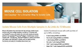
 EasySep™小鼠TIL(CD45)正选试剂盒


 实验方案Optimizing Delivery Efficiency with Fluorescent Dextran Using the CellPore™ Transfection System


 沪公网安备31010102008431号