Naramura M et al. (SEP 2010)
Proceedings of the National Academy of Sciences of the United States of America 107 37 16274--9
Rapidly fatal myeloproliferative disorders in mice with deletion of Casitas B-cell lymphoma (Cbl) and Cbl-b in hematopoietic stem cells.
Casitas B-cell lymphoma (Cbl)-family E3 ubiquitin ligases are negative regulators of tyrosine kinase signaling. Recent work has revealed a critical role of Cbl in the maintenance of hematopoietic stem cell (HSC) homeostasis,and mutations in CBL have been identified in myeloid malignancies. Here we show that,in contrast to Cbl or Cbl-b single-deficient mice,concurrent loss of Cbl and Cbl-b in the HSC compartment leads to an early-onset lethal myeloproliferative disease in mice. Cbl,Cbl-b double-deficient bone marrow cells are hypersensitive to cytokines,and show altered biochemical response to thrombopoietin. Thus,Cbl and Cbl-b play redundant but essential roles in HSC regulation,whose breakdown leads to hematological abnormalities that phenocopy crucial aspects of mutant Cbl-driven human myeloid malignancies.
View Publication
产品号#:
03234
产品名:
MethoCult™ M3234
Wang J et al. (SEP 2010)
Proceedings of the National Academy of Sciences of the United States of America 107 37 16131--6
CCAAT/enhancer binding protein delta (C/EBPdelta, CEBPD)-mediated nuclear import of FANCD2 by IPO4 augments cellular response to DNA damage.
Maintenance of genomic integrity is an essential cellular function. We previously reported that the transcription factor and tumor suppressor CCAAT/enhancer binding protein δ (C/EBPδ,CEBPD; also known as NFIL-6β") promotes genomic stability. However�
View Publication
产品号#:
03434
03444
产品名:
MethoCult™ GF M3434
MethoCult™ GF M3434
Tzeng Y-S et al. (JAN 2011)
Blood 117 2 429--39
Loss of Cxcl12/Sdf-1 in adult mice decreases the quiescent state of hematopoietic stem/progenitor cells and alters the pattern of hematopoietic regeneration after myelosuppression.
The C-X-C-type chemokine Cxcl12,also known as stromal cell-derived factor-1,plays a critical role in hematopoiesis during fetal development. However,the functional requirement of Cxcl12 in the adult hematopoietic stem/progenitor cell (HSPC) regulation was still unclear. In this report,we developed a murine Cxcl12 conditional deletion model in which the target gene can be deleted at the adult stage. We found that loss of stroma-secreted Cxcl12 in the adult led to expansion of the HSPC population as well as a reduction in long-term quiescent stem cells. In Cxcl12-deficient bone marrow,HSPCs were absent along the endosteal surface,and blood cell regeneration occurred predominantly in the perisinusoidal space after 5-fluorouracil myelosuppression challenge. Our results indicate that Cxcl12 is required for HSPC homeostasis regulation and is an important factor for osteoblastic niche organization in adult stage bone marrow.
View Publication
产品号#:
03434
03444
产品名:
MethoCult™ GF M3434
MethoCult™ GF M3434
Xiao W et al. (DEC 2010)
Blood 116 26 6003--13
Lyn- and PLC-beta3-dependent regulation of SHP-1 phosphorylation controls Stat5 activity and myelomonocytic leukemia-like disease.
Hyperactivation of the transcription factor Stat5 leads to various leukemias. Stat5 activity is regulated by the protein phosphatase SHP-1 in a phospholipase C (PLC)-β3-dependent manner. Thus,PLC-β3-deficient mice develop myeloproliferative neoplasm,like Lyn (Src family kinase)- deficient mice. Here we show that Lyn/PLC-β3 doubly deficient lyn(-/-);PLC-β3(-/-) mice develop a Stat5-dependent,fatal myelodysplastic/myeloproliferative neoplasm,similar to human chronic myelomonocytic leukemia (CMML). In hematopoietic stem cells of lyn(-/-);PLC-β3(-/-) mice that cause the CMML-like disease,phosphorylation of SHP-1 at Tyr(536) and Tyr(564) is abrogated,resulting in reduced phosphatase activity and constitutive activation of Stat5. Furthermore,SHP-1 phosphorylation at Tyr(564) by Lyn is indispensable for maximal phosphatase activity and for suppression of the CMML-like disease in these mice. On the other hand,Tyr(536) in SHP-1 can be phosphorylated by Lyn and another kinase(s) and is necessary for efficient interaction with Stat5. Therefore,we identify a novel Lyn/PLC-β3-mediated regulatory mechanism of SHP-1 and Stat5 activities.
View Publication
产品号#:
03134
产品名:
MethoCult™ M3134
Lidonnici MR et al. (OCT 2010)
Cancer research 70 20 7949--59
Expression of the transcriptional repressor Gfi-1 is regulated by C/EBPalpha and is involved in its proliferation and colony formation-inhibitory effects in p210BCR/ABL-expressing cells.
Ectopic expression of CAAT/enhancer binding protein α (C/EBPα) in p210BCR/ABL-expressing cells induces granulocytic differentiation,inhibits proliferation,and suppresses leukemogenesis. To dissect the molecular mechanisms underlying these biological effects,C/EBPα-regulated genes were identified by microarray analysis in 32D-p210BCR/ABL cells. One of the genes whose expression was activated by C/EBPα in a DNA binding-dependent manner in BCR/ABL-expressing cells is the transcriptional repressor Gfi-1. We show here that C/EBPα interacts with a functional C/EBP binding site in the Gfi-1 5'-flanking region and enhances the promoter activity of Gfi-1. Moreover,in K562 cells,RNA interference-mediated downregulation of Gfi-1 expression partially rescued the proliferation-inhibitory but not the differentiation-inducing effect of C/EBPα. Ectopic expression of wild-type Gfi-1,but not of a transcriptional repressor mutant (Gfi-1P2A),inhibited proliferation and markedly suppressed colony formation but did not induce granulocytic differentiation of BCR/ABL-expressing cells. By contrast,Gfi-1 short hairpin RNA-tranduced CD34(+) chronic myeloid leukemia cells were markedly more clonogenic than the scramble-transduced counterpart. Together,these studies indicate that Gfi-1 is a direct target of C/EBPα required for its proliferation and survival-inhibitory effects in BCR/ABL-expressing cells.
View Publication
产品号#:
02690
09600
09650
产品名:
StemSpan™ CC100
StemSpan™ SFEM
StemSpan™ SFEM
Ma ACH et al. (DEC 2010)
Leukemia 24 12 2090--9
A DEAB-sensitive aldehyde dehydrogenase regulates hematopoietic stem and progenitor cells development during primitive hematopoiesis in zebrafish embryos.
Although aldehyde dehydrogenase (ALDH) activity has become a surrogate of hematopoietic stem and progenitor cells (HSPCs),its function during hematopoiesis was unclear. Here,we examined its role in zebrafish hematopoiesis based on pharmacological inhibition and morpholino (MO) knockdown. Zebrafish embryos were treated with diethylaminobenzaldehyde (DEAB,1 μmol/l) between 0- and 48 hour-post-fertilization (hpf). MOs targeting aldhs were injected between 1 and 4-cell stage. The effects on hematopoiesis were evaluated at different stages. DEAB treatment between 0 and 18 hpf increased gene expression associated with HSPC (scl,lmo2),erythropoiesis (gata1,α- and β-eHb) and myelopoiesis (spi1) as well as gfp(+) cells in dissociated Tg(gata1:gfp) embryos. The effects were ameliorated by all-trans retinoic acid (1 nmol/l). Definitive hematopoiesis and the erythromyeloid precursors were unaffected. In all,14 out of 15 zebrafish aldhs were detectable by reverse transcription PCR in 18 hpf embryos,of which only aldh1a2 and aldh16a1 were expressed in sites pertinent to hematopoiesis. Molecular targeting by MOs was demonstrated for 15 aldhs,but none of them,even in combined aldh1a2 and aldh1a3 knockdown,recapitulated the hematopoietic expansion in DEAB-treated embryos. In conclusion,DEAB expands HSPC population during primitive hematopoiesis through inhibition of aldh and retinoic acid synthesis. The specific aldh isoform(s) remains to be determined.
View Publication
产品号#:
01700
01705
01702
产品名:
ALDEFLUOR™ 试剂盒
ALDEFLUOR™ DEAB试剂, 1.5 mM, 1 mL
ALDEFLUOR™检测缓冲液
Mahdipour E et al. (JAN 2011)
Blood 117 3 815--26
Hoxa3 promotes the differentiation of hematopoietic progenitor cells into proangiogenic Gr-1+CD11b+ myeloid cells.
Injury induces the recruitment of bone marrow-derived cells (BMDCs) that contribute to the repair and regeneration process. The behavior of BMDCs in injured tissue has a profound effect on repair,but the regulation of BMDC behavior is poorly understood. Aberrant recruitment/retention of these cells in wounds of diabetic patients and animal models is associated with chronic inflammation and impaired healing. BMD Gr-1(+)CD11b(+) cells function as immune suppressor cells and contribute significantly to tumor-induced neovascularization. Here we report that Gr-1(+)CD11b(+) cells also contribute to injury-induced neovascularization,but show altered recruitment/retention kinetics in the diabetic environment. Moreover,diabetic-derived Gr-1(+)CD11b(+) cells fail to stimulate neovascularization in vivo and have aberrant proliferative,chemotaxis,adhesion,and differentiation potential. Previously we demonstrated that gene transfer of HOXA3 to wounds of diabetic mice is taken up by and expressed by recruited BMDCs. This is associated with a suppressed inflammatory response,enhanced neovascularization,and accelerated wound healing. Here we show that sustained expression of Hoxa3 in diabetic-derived BMD Gr-1(+)CD11b(+) cells reverses their diabetic phenotype. These findings demonstrate that manipulation of adult stem/progenitor cells ex vivo could be used as a potential therapy in patients with impaired wound healing.
View Publication
产品号#:
03434
03444
产品名:
MethoCult™ GF M3434
MethoCult™ GF M3434
Stutz MD et al. (DEC 2017)
Cell death and differentiation
Necroptotic signaling is primed in Mycobacterium tuberculosis-infected macrophages, but its pathophysiological consequence in disease is restricted.
Mixed lineage kinase domain-like (MLKL)-dependent necroptosis is thought to be implicated in the death of mycobacteria-infected macrophages,reportedly allowing escape and dissemination of the microorganism. Given the consequent interest in developing inhibitors of necroptosis to treat Mycobacterium tuberculosis (Mtb) infection,we used human pharmacologic and murine genetic models to definitively establish the pathophysiological role of necroptosis in Mtb infection. We observed that Mtb infection of macrophages remodeled the intracellular signaling landscape by upregulating MLKL,TNFR1,and ZBP1,whilst downregulating cIAP1,thereby establishing a strong pro-necroptotic milieu. However,blocking necroptosis either by deleting Mlkl or inhibiting RIPK1 had no effect on the survival of infected human or murine macrophages. Consistent with this,MLKL-deficiency or treatment of humanized mice with the RIPK1 inhibitor Nec-1s did not impact on disease outcomes in vivo,with mice displaying lung histopathology and bacterial burdens indistinguishable from controls. Therefore,although the necroptotic pathway is primed by Mtb infection,macrophage necroptosis is ultimately restricted to mitigate disease pathogenesis. We identified cFLIP upregulation that may promote caspase 8-mediated degradation of CYLD,and other necrosome components,as a possible mechanism abrogating Mtb's capacity to coopt necroptotic signaling. Variability in the capacity of these mechanisms to interfere with necroptosis may influence disease severity and could explain the heterogeneity of Mtb infection and disease.
View Publication

 EasySep™小鼠TIL(CD45)正选试剂盒


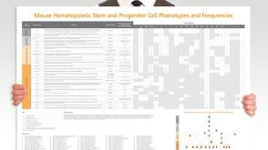 挂图Mouse Hematopoietic Stem and Progenitor Cell Phenotyping Overview of mouse HSPC subset surface markers and frequencies


 沪公网安备31010102008431号