Progenitor cell dose determines the pace and completeness of engraftment in a xenograft model for cord blood transplantation.
Two critical concerns in clinical cord blood transplantation are the initial time to engraftment and the subsequent restoration of immune function. These studies measured the impact of progenitor cell dose on both the pace and strength of hematopoietic reconstitution by transplanting nonobese diabetic/severe combined immunodeficiency/interleukin-2 receptor-gamma-null (NSγ) mice with lineage-depleted aldehyde dehydrogenase-bright CD34(+) human cord blood progenitors. The progress of each transplant was monitored over an extended time course by repeatedly analyzing the peripheral blood for human hematopoietic cells. In vivo human hematopoietic development was complete. After long-term transplantation assays (≥ 19 weeks),human T-cell development was documented within multiple tissues in 16 of 32 NSγ mice. Human T-cell differentiation was active within NSγ thymuses,as documented by the presence of CD4(+) CD8(+) T-cell progenitors as well as T-cell receptor excision circles. It is important to note that although myeloid and B-cell engraftment was detected as early as 4 weeks after transplantation,human T-cell development was exclusively late onset. High progenitor cell doses were associated with a robust human hematopoietic chimerism that accelerated both initial time to engraftment and subsequent T-cell development. At lower progenitor cell doses,the chimerism was weak and the human hematopoietic lineage development was frequently incomplete.
View Publication
产品号#:
01700
01705
01701
01702
14056
14066
28600
19056
19056RF
19756
19756RF
产品名:
ALDEFLUOR™ 试剂盒
ALDEFLUOR™ DEAB试剂, 1.5 mM, 1 mL
ALDEFLUOR™检测缓冲液
L-Calc™有限稀释软件
Ryan MA et al. (OCT 2010)
Nature medicine 16 10 1141--6
Mobilization of hematopoietic stem and progenitor cells (HSPCs) from bone marrow into peripheral blood by the cytokine granulocyte colony-stimulating factor (G-CSF) has become the preferred source of HSPCs for stem cell transplants. However,G-CSF fails to mobilize sufficient numbers of stem cells in up to 10% of donors,precluding autologous transplantation in those donors or substantially delaying transplant recovery time. Consequently,new regimens are needed to increase the number of stem cells in peripheral blood upon mobilization. Using a forward genetic approach in mice,we mapped the gene encoding the epidermal growth factor receptor (Egfr) to a genetic region modifying G-CSF-mediated HSPC mobilization. Amounts of EGFR in HSPCs inversely correlated with the cells' ability to be mobilized by G-CSF,implying a negative role for EGFR signaling in mobilization. In combination with G-CSF treatment,genetic reduction of EGFR activity in HSPCs (in waved-2 mutant mice) or treatment with the EGFR inhibitor erlotinib increased mobilization. Increased mobilization due to suppression of EGFR activity correlated with reduced activity of cell division control protein-42 (Cdc42),and genetic Cdc42 deficiency in vivo also enhanced G-CSF-induced mobilization. Our findings reveal a previously unknown signaling pathway regulating stem cell mobilization and provide a new pharmacological approach for improving HSPC mobilization and thereby transplantation outcomes.
View Publication
产品号#:
03234
产品名:
MethoCult™ M3234
Park S-W et al. (DEC 2010)
Blood 116 25 5762--72
Efficient differentiation of human pluripotent stem cells into functional CD34+ progenitor cells by combined modulation of the MEK/ERK and BMP4 signaling pathways.
Differentiation of human pluripotent stem cells (hPSCs) into functional cell types is a crucial step in cell therapy. In the present study,we demonstrate that functional CD34(+) progenitor cells can be efficiently produced from human embryonic stem cells (hESCs) and induced pluripotent stem cells (hiPSCs) by combined modulation of 2 signaling pathways. A higher proportion of CD34(+) cells (∼ 20%) could be derived from hPSCs by inhibition of mitogen-activated protein kinase (MAPK) extracellular signal-regulated protein kinase (MEK)/extracellular signal-regulated kinase (ERK) signaling and activation of bone morphogenic protein-4 (BMP4) signaling. hPSC-derived CD34(+) progenitor cells further developed to endothelial and smooth muscle cells with functionality. Moreover,they contributed directly to neovasculogenesis in ischemic mouse hind limbs,thereby resulting in improved blood perfusion and limb salvage. Our results suggest that combined modulation of signaling pathways may be an efficient means of differentiating hPSCs into functional CD34(+) progenitor cells.
View Publication

 EasySep™小鼠TIL(CD45)正选试剂盒


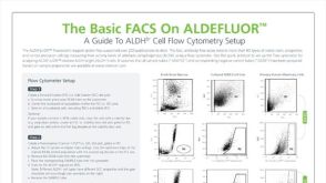 技术公告The Basic FACS on ALDEFLUOR™: The Quick Guide to Flow Cytometry
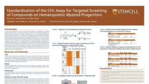
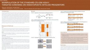 科学海报Manipulation of the Standard CFU-GM Assay Targeted Screening of Hematopoietic Myeloid Progenitors
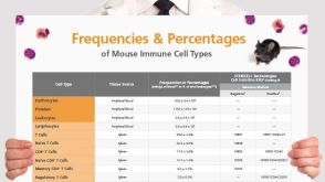 挂图Frequencies and Percentages of Mouse Immune Cell Types List of the frequencies of over 25 immune cell types in C57BL/6 mice


 沪公网安备31010102008431号