Scott Allen, PhD
Seeking Metabolic Therapies for an Incurable Neurodegenerative Disease
研究方向:
干细胞生物学,神经科学
Li L et al. (JUL 2010)
The Journal of neuroscience : the official journal of the Society for Neuroscience 30 27 9038--50
Endogenous interferon gamma directly regulates neural precursors in the non-inflammatory brain.
Although a number of growth factors have been shown to be involved in neurogenesis,the role of inflammatory cytokines remains relatively unexplored in the normal brain. Here we investigated the effect of interferon gamma (IFNgamma) in the regulation of neural precursor (NP) activity in both the developing and the adult mouse brain. Exogenous IFNgamma inhibited neurosphere formation from the wild-type neonatal and adult subventricular zone (SVZ). More importantly,however,these effects were mirrored in vivo,with mutant mice lacking endogenous IFNgamma displaying enhanced neurogenesis,as demonstrated by an increase in proliferative bromodeoxyuridine-labeled cells in the SVZ and an increased percentage of newborn neurons in the olfactory bulb. Furthermore,NPs isolated from IFNgamma null mice exhibited an increase in self-renewal ability and in the capacity to produce differentiated neurons and oligodendrocytes. These effects resulted from the direct action of IFNgamma on the NPs,as determined by single-cell assays and the fact that nearly all the neurospheres were derived from cells positive for major histocompatibility complex class I antigen,a downstream marker of IFNgamma-mediated activation. Moreover,the inhibitory effect was ameliorated in the presence of SVZ-derived microglia,with their removal resulting in almost complete inhibition of NP proliferation. Interestingly,in contrast to the results obtained in the adult,exogenous IFNgamma treatment stimulated neurosphere formation from the embryonic brain,an effect that was mediated by sonic hedgehog. Together these findings provide the first direct evidence that IFNgamma acts as a regulator of the active NP pool in the non-inflammatory brain.
View Publication
产品号#:
05700
05701
05702
产品名:
NeuroCult™ 基础培养基(小鼠和大鼠)
NeuroCult™ 扩增添加物(小鼠和大鼠)
NeuroCult™扩增试剂盒(小鼠和大鼠)
Huat T et al. (JUL 2014)
BMC Neuroscience 15 1 91
IGF-1 enhances cell proliferation and survival during early differentiation of mesenchymal stem cells to neural progenitor-like cells
BACKGROUND There has been increasing interest recently in the plasticity of mesenchymal stem cells (MSCs) and their potential to differentiate into neural lineages. To unravel the roles and effects of different growth factors in the differentiation of MSCs into neural lineages,we have differentiated MSCs into neural lineages using different combinations of growth factors. Based on previous studies of the roles of insulin-like growth factor 1 (IGF-1) in neural stem cell isolation in the laboratory,we hypothesized that IGF-1 can enhance proliferation and reduce apoptosis in neural progenitor-like cells (NPCs) during differentiation of MSCs into NCPs.We induced MSCs differentiation under four different combinations of growth factors: (A) EGF%+%bFGF,(B) EGF%+%bFGF%+%IGF-1,(C) EGF%+%bFGF%+%LIF,(D) EGF%+%bFGF%+%BDNF,and (E) without growth factors,as a negative control. The neurospheres formed were characterized by immunofluorescence staining against nestin,and the expression was measured by flow cytometry. Cell proliferation and apoptosis were also studied by MTS and Annexin V assay,respectively,at three different time intervals (24 hr,3 days,and 5 days). The neurospheres formed in the four groups were then terminally differentiated into neuron and glial cells. RESULTS The four derived NPCs showed a significantly higher expression of nestin than was shown by the negative control. Among the groups treated with growth factors,NPCs treated with IGF-1 showed the highest expression of nestin. Furthermore,NPCs derived using IGF-1 exhibited the highest cell proliferation and cell survival among the treated groups. The NPCs derived from IGF-1 treatment also resulted in a better yield after the terminal differentiation into neurons and glial cells than that of the other treated groups. CONCLUSIONS Our results suggested that IGF-1 has a crucial role in the differentiation of MSCs into neuronal lineage by enhancing the proliferation and reducing the apoptosis in the NPCs. This information will be beneficial in the long run for improving both cell-based and cell-free therapy for neurodegenerative diseases.
View Publication
产品号#:
05771
产品名:
Abeysinghe HCS et al. (SEP 2015)
Stem cell research & therapy 6 1 186
Pre-differentiation of human neural stem cells into GABAergic neurons prior to transplant results in greater repopulation of the damaged brain and accelerates functional recovery after transient ischemic stroke.
INTRODUCTION Despite attempts to prevent brain injury during the hyperacute phase of stroke,most sufferers end up with significant neuronal loss and functional deficits. The use of cell-based therapies to recover the injured brain offers new hope. In the current study,we employed human neural stem cells (hNSCs) isolated from subventricular zone (SVZ),and directed their differentiation into GABAergic neurons followed by transplantation to ischemic brain. METHODS Pre-differentiated GABAergic neurons,undifferentiated SVZ-hNSCs or media alone were stereotaxically transplanted into the rat brain (n=7/group) 7 days after endothelin-1 induced stroke. Neurological outcome was assessed by neurological deficit scores and the cylinder test. Transplanted cell survival,cellular phenotype and maturation were assessed using immunohistochemistry and confocal microscopy. RESULTS Behavioral assessments revealed accelerated improvements in motor function 7 days post-transplant in rats treated with pre-differentiated GABAergic cells in comparison to media alone and undifferentiated hNSC treated groups. Histopathology 28 days-post transplant indicated that pre-differentiated cells maintained their GABAergic neuronal phenotype,showed evidence of synaptogenesis and up-regulated expression of both GABA and calcium signaling proteins associated with neurotransmission. Rats treated with pre-differentiated cells also showed increased neurogenic activity within the SVZ at 28 days,suggesting an additional trophic role of these GABAergic cells. In contrast,undifferentiated SVZ-hNSCs predominantly differentiated into GFAP-positive astrocytes and appeared to be incorporated into the glial scar. CONCLUSION Our study is the first to show enhanced exogenous repopulation of a neuronal phenotype after stroke using techniques aimed at GABAergic cell induction prior to delivery that resulted in accelerated and improved functional recovery.
View Publication
产品号#:
05750
05751
产品名:
NeuroCult™ NS-A 基础培养基(人)
NeuroCult™ NS-A 扩增试剂盒(人)
Alison MR et al. (DEC 2010)
The Journal of pathology 222 4 335--44
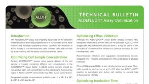
Finding cancer stem cells: are aldehyde dehydrogenases fit for purpose?
Despite many years of intensive effort,there is surprisingly little consensus on the most suitable markers with which to locate and isolate stem cells from adult tissues. By comparison,the study of cancer stem cells is still in its infancy; so,unsurprisingly,there is great uncertainty as to the identity of these cells. Stem cell markers can be broadly categorized into molecular determinants of self-renewal,clonogenicity,multipotentiality,adherence to the niche,and longevity. This review assesses the utility of recognizing cancer stem cells by virtue of high expression of aldehyde dehydrogenases (ALDHs),probably significant determinants of cell survival through their ability to detoxify many potentially cytotoxic molecules,and contributing to drug resistance. Antibodies are available against the ALDH enzyme family,but the vast majority of studies have used cell sorting techniques to enrich for cells expressing these enzymes. Live cells expressing high ALDH activity are usually identified by the ALDEFLUOR kit and sorted by fluorescence activated cell sorting (FACS). For many human tumours,but notably breast cancer,cell selection based upon ALDH activity appears to be a useful marker for enriching for cells with tumour-initiating activity (presumed cancer stem cells) in immunodeficient mice,and indeed the frequency of so-called ALDH(bri) cells in many tumours can be an independent prognostic indicator.
View Publication
alpha1-Adrenergic receptors regulate neurogenesis and gliogenesis.
The understanding of the function of alpha(1)-adrenergic receptors in the brain has been limited due to a lack of specific ligands and antibodies. We circumvented this problem by using transgenic mice engineered to overexpress either wild-type receptor tagged with enhanced green fluorescent protein or constitutively active mutant alpha(1)-adrenergic receptor subtypes in tissues in which they are normally expressed. We identified intriguing alpha(1A)-adrenergic receptor subtype-expressing cells with a migratory morphology in the adult subventricular zone that coexpressed markers of neural stem cell and/or progenitors. Incorporation of 5-bromo-2-deoxyuridine in vivo increased in neurogenic areas in adult alpha(1A)-adrenergic receptor transgenic mice or normal mice given the alpha(1A)-adrenergic receptor-selective agonist,cirazoline. Neonatal neurospheres isolated from normal mice expressed a mixture of alpha(1)-adrenergic receptor subtypes,and stimulation of these receptors resulted in increased expression of the alpha(1B)-adrenergic receptor subtype,proneural basic helix-loop-helix transcription factors,and the differentiation and migration of neuronal progenitors for catecholaminergic neurons and interneurons. alpha(1)-Adrenergic receptor stimulation increased the apoptosis of astrocytes and regulated survival of neonatal neurons through phosphatidylinositol 3-kinase signaling. However,in adult normal neurospheres,alpha(1)-adrenergic receptor stimulation increased the expression of glial markers at the expense of neuronal differentiation. In vivo,S100-positive glial and betaIII tubulin neuronal progenitors colocalized with either alpha(1)-adrenergic receptor subtype in the olfactory bulb. Our results indicate that alpha(1)-adrenergic receptors can regulate both neurogenesis and gliogenesis that may be developmentally dependent. Our findings may lead to new therapies to treat neurodegenerative diseases.
View Publication

 EasySep™小鼠TIL(CD45)正选试剂盒


 科学海报Single-Cell RNA Sequencing Analysis of Regionally Patterned Human Pluripotent Stem Cell-Derived Neural Organoids
 专家访谈Scott Allen, PhD Seeking Metabolic Therapies for an Incurable Neurodegenerative Disease

 沪公网安备31010102008431号