
 EasySep™小鼠TIL(CD45)正选试剂盒


技术资料
-
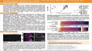 科学海报Using Human Pluripotent Stem Cell-Derived Microglia As Models For Neurological Disease Research
Conference:
FENS 2020
发布日期: 07/24/2020 -
-

-
-
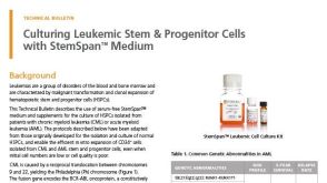 技术公告Culturing Leukemic Stem & Progenitor Cells with StemSpan™ Medium
细胞类型:
白血病/淋巴瘤细胞
发布日期: 11/14/2019


 沪公网安备31010102008431号