Finstad SL et al. (JUL 2007)
Journal of virology 81 13 7274--9
Diminished potential for B-lymphoid differentiation after murine leukemia virus infection in vivo and in EML hematopoietic progenitor cells.
Infection with a recombinant murine-feline gammaretrovirus,MoFe2,or with the parent virus,Moloney murine leukemia virus,caused significant reduction in B-lymphoid differentiation of bone marrow at 2 to 8 weeks postinfection. The suppression was selective,in that myeloid potential was significantly increased by infection. Analysis of cell surface markers and immunoglobulin H gene rearrangements in an in vitro model demonstrated normal B-lymphoid differentiation after infection but significantly reduced viability of differentiating cells. This reduction in viability may confer a selective advantage on undifferentiated lymphoid progenitors in the bone marrow of gammaretrovirus-infected animals and thereby contribute to the establishment of a premalignant state.
View Publication
产品号#:
03630
03434
03444
产品名:
MethoCult™ M3630
MethoCult™ GF M3434
MethoCult™ GF M3434
Rawat VPS et al. (JAN 2008)
Blood 111 1 309--19
Overexpression of CDX2 perturbs HOX gene expression in murine progenitors depending on its N-terminal domain and is closely correlated with deregulated HOX gene expression in human acute myeloid leukemia.
The mechanisms underlying deregulation of HOX gene expression in AML are poorly understood. The ParaHox gene CDX2 was shown to act as positive upstream regulator of several HOX genes. In this study,constitutive expression of Cdx2 caused perturbation of leukemogenic Hox genes such as Hoxa10 and Hoxb8 in murine hematopoietic progenitors. Deletion of the N-terminal domain of Cdx2 abrogated its ability to perturb Hox gene expression and to cause acute myeloid leukemia (AML) in mice. In contrast inactivation of the putative Pbx interacting site of Cdx2 did not change the leukemogenic potential of the gene. In an analysis of 115 patients with AML,expression levels of CDX2 were closely correlated with deregulated HOX gene expression. Patients with normal karyotype showed a 14-fold higher expression of CDX2 and deregulated HOX gene expression compared with patients with chromosomal translocations such as t(8:21) or t(15;17). All patients with AML with normal karyotype tested were negative for CDX1 and CDX4 expression. These data link the leukemogenic potential of Cdx2 to its ability to dysregulate Hox genes. They furthermore correlate the level of CDX2 expression with HOX gene expression in human AML and support a potential role of CDX2 in the development of human AML with aberrant Hox gene expression.
View Publication
Pei S et al. (NOV 2013)
The Journal of biological chemistry 288 47 33542--58
Targeting aberrant glutathione metabolism to eradicate human acute myelogenous leukemia cells.
The development of strategies to eradicate primary human acute myelogenous leukemia (AML) cells is a major challenge to the leukemia research field. In particular,primitive leukemia cells,often termed leukemia stem cells,are typically refractory to many forms of therapy. To investigate improved strategies for targeting of human AML cells we compared the molecular mechanisms regulating oxidative state in primitive (CD34(+)) leukemic versus normal specimens. Our data indicate that CD34(+) AML cells have elevated expression of multiple glutathione pathway regulatory proteins,presumably as a mechanism to compensate for increased oxidative stress in leukemic cells. Consistent with this observation,CD34(+) AML cells have lower levels of reduced glutathione and increased levels of oxidized glutathione compared with normal CD34(+) cells. These findings led us to hypothesize that AML cells will be hypersensitive to inhibition of glutathione metabolism. To test this premise,we identified compounds such as parthenolide (PTL) or piperlongumine that induce almost complete glutathione depletion and severe cell death in CD34(+) AML cells. Importantly,these compounds only induce limited and transient glutathione depletion as well as significantly less toxicity in normal CD34(+) cells. We further determined that PTL perturbs glutathione homeostasis by a multifactorial mechanism,which includes inhibiting key glutathione metabolic enzymes (GCLC and GPX1),as well as direct depletion of glutathione. These findings demonstrate that primitive leukemia cells are uniquely sensitive to agents that target aberrant glutathione metabolism,an intrinsic property of primary human AML cells.
View Publication
Mahbub AA et al. (DEC 2013)
Anti-cancer agents in medicinal chemistry 13 10 1601--13
Differential effects of polyphenols on proliferation and apoptosis in human myeloid and lymphoid leukemia cell lines.
BACKGROUND Mortality rates for leukemia are high despite considerable improvements in treatment. Since polyphenols exert pro-apoptotic effects in solid tumors,our study investigated the effects of polyphenols in haematological malignancies. The effect of eight polyphenols (quercetin,chrysin,apigenin,emodin,aloe-emodin,rhein,cis-stilbene and trans-stilbene) were studied on cell proliferation,cell cycle and apoptosis in four lymphoid and four myeloid leukemic cells lines,together with normal haematopoietic control cells. METHODS Cellular proliferation was measured by CellTiter-Glo(®) luminescent assay; and cell cycle arrest was assessed using flow cytometry of propidium iodide stained cells. Apoptosis was investigated by caspase-3 activity assay using flow cytometry and apoptotic morphology was confirmed by Hoescht 33342 staining. RESULTS Emodin,quercetin,and cis-stilbene were the most effective polyphenols at decreasing cell viability (IC50 values of 5-22 μM,8-33 μM,and 25-85 μM respectively) and inducing apoptosis (AP50 values (the concentration which 50% of cells undergo apoptosis) of 2-27 μM,19-50 μM,and 8-50 μM respectively). Generally,lymphoid cell lines were more sensitive to polyphenol treatment compared to myeloid cell lines,however the most resistant myeloid (KG-1a and K562) cell lines were still found to respond to emodin and quercetin treatment at low micromolar levels. Non-tumor cells were less sensitive to all polyphenols compared to the leukemia cells. CONCLUSIONS These findings suggest that polyphenols have anti-tumor activity against leukemia cells with differential effects. Importantly,the differential sensitivity of emodin,quercetin,and cis-stilbene between leukemia and normal cells suggests that polyphenols are potential therapeutic agents for leukemia.
View Publication
产品号#:
70008
70008.1
70008.2
70008.3
70008.4
70008.5
70008.6
200-0002
200-0001
200-0000
产品名:
冻存的人脐带血CD34+细胞
冻存的人脐带血CD34+细胞
冻存的人脐带血CD34+细胞
冻存的人脐带血CD34+细胞
冻存的人脐带血CD34+细胞
冻存的人脐带血CD34+细胞
冻存的人脐带血CD34+细胞
冻存的人脐带血CD34+细胞
冻存的人脐带血CD34+细胞
Elliott S et al. (JUL 2013)
PloS one 8 7 e68083
Epo receptors are not detectable in primary human tumor tissue samples.
Erythropoietin (Epo) is a cytokine that binds and activates an Epo receptor (EpoR) expressed on the surface of erythroid progenitor cells to promote erythropoiesis. While early studies suggested EpoR transcripts were expressed exclusively in the erythroid compartment,low-level EpoR transcripts were detected in nonhematopoietic tissues and tumor cell lines using sensitive RT-PCR methods. However due to the widespread use of nonspecific anti-EpoR antibodies there are conflicting data on EpoR protein expression. In tumor cell lines and normal human tissues examined with a specific and sensitive monoclonal antibody to human EpoR (A82),little/no EpoR protein was detected and it was not functional. In contrast,EpoR protein was reportedly detectable in a breast tumor cell line (MCF-7) and breast cancer tissues with an anti-EpoR polyclonal antibody (M-20),and functional responses to rHuEpo were reported with MCF-7 cells. In another study,a functional response was reported with the lung tumor cell line (NCI-H838) at physiological levels of rHuEpo. However,the specificity of M-20 is in question and the absence of appropriate negative controls raise questions about possible false-positive effects. Here we show that with A82,no EpoR protein was detectable in normal human and matching cancer tissues from breast,lung,colon,ovary and skin with little/no EpoR in MCF-7 and most other breast and lung tumor cell lines. We show further that M-20 provides false positive staining with tissues and it binds to a non-EpoR protein that migrates at the same size as EpoR with MCF-7 lysates. EpoR protein was detectable with NCI-H838 cells,but no rHuEpo-induced phosphorylation of AKT,STAT3,pS6RP or STAT5 was observed suggesting the EpoR was not functional. Taken together these results raise questions about the hypothesis that most tumors express high levels of functional EpoR protein.
View Publication
Marcato P et al. (MAY 2011)
Cell cycle (Georgetown,Tex.) 10 9 1378--84
Aldehyde dehydrogenase: its role as a cancer stem cell marker comes down to the specific isoform.
Recent evidence suggests that enhanced aldehyde dehydrogenase (ALDH) activity is a hallmark of cancer stem cells (CSC) measurable by the aldefluor assay. ALDH1A1,one of 19 ALDH isoforms expressed in humans,was generally believed to be responsible for the ALDH activity of CSCs. More recently,experiments with murine hematopoietic stem cells,murine progenitor pancreatic cells,and human breast CSCs indicate that other ALDH isoforms,particularly ALDH1A3,significantly contribute to aldefluor positivity,which may be tissue and cancer specific. Therefore,potential prognostic application involving the use of CSC prevalence in tumor tissue to predict patient outcome requires the identification and quantification of specific ALDH isoforms. Herein we review the suggested roles of ALDH in CSC biology and the immunohistological studies testing the potential application of ALDH isoforms as novel cancer prognostic indicators.
View Publication

 EasySep™小鼠TIL(CD45)正选试剂盒


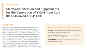 技术公告StemSpan™ Medium and Supplements for the Generation of T Cells from Cord Blood-Derived CD34+ Cells


 沪公网安备31010102008431号