Cremona CA and Lloyd AC (SEP 2009)
Journal of cell science 122 Pt 18 3272--81
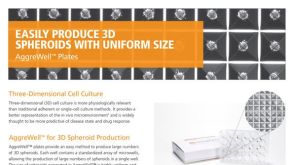
Loss of anchorage in checkpoint-deficient cells increases genomic instability and promotes oncogenic transformation.
Mammalian cells generally require both mitogens and anchorage signals in order to proliferate. An important characteristic of many tumour cells is that they have lost this anchorage-dependent cell-cycle checkpoint,allowing them to proliferate without signals provided by their normal microenvironment. In the absence of anchorage signals from the extracellular matrix,many cell types arrest cell-cycle progression in G1 phase as a result of Rb-dependent checkpoints. However,despite inactivation of p53 and Rb proteins,SV40LT-expressing cells retain anchorage dependency,suggesting the presence of an uncharacterised cell-cycle checkpoint,which can be overridden by coexpression of oncogenic Ras. We report here that,although cyclin-CDK complexes persisted in suspension,proliferation was inhibited in LT-expressing cells by the CDK inhibitor p27(Kip1) (p27). Interestingly,this did not induce a stable arrest,but aberrant cell-cycle progression associated with stalled DNA replication,rereplication and chromosomal instability,which was sufficient to increase the frequency of oncogenic transformation. These results firstly indicate loss of anchorage in Rb- and p53-deficient cells as a novel mechanism for promotion of genomic instability; secondly suggest that anchorage checkpoints that protect normal cells from inappropriate proliferation act deleteriously in Rb- and p53-deficient cells to promote tumourigenesis; and thirdly indicate caution in the use of CDK inhibitors for cancer treatment.
View Publication
Zhao Q et al. (JAN 2015)
Proceedings of the National Academy of Sciences of the United States of America 112 2 530--535
MSCs derived from iPSCs with a modified protocol are tumor-tropic but have much less potential to promote tumors than bone marrow MSCs.
Mesenchymal stem or stromal cells (MSCs) have many potential therapeutic applications including therapies for cancers and tissue damages caused by cancers or radical cancer treatments. However,tissue-derived MSCs such as bone marrow MSCs (BM-MSCs) may promote cancer progression and have considerable donor variations and limited expandability. These issues hinder the potential applications of MSCs,especially those in cancer patients. To circumvent these issues,we derived MSCs from transgene-free human induced pluripotent stem cells (iPSCs) efficiently with a modified protocol that eliminated the need of flow cytometric sorting. Our iPSC-derived MSCs were readily expandable,but still underwent senescence after prolonged culture and did not form teratomas. These iPSC-derived MSCs homed to cancers with efficiencies similar to BM-MSCs but were much less prone than BM-MSCs to promote the epithelial-mesenchymal transition,invasion,stemness,and growth of cancer cells. The observations were probably explained by the much lower expression of receptors for interleukin-1 and TGFβ,downstream protumor factors,and hyaluronan and its cofactor TSG6,which all contribute to the protumor effects of BM-MSCs. The data suggest that iPSC-derived MSCs prepared with the modified protocol are a safer and better alternative to BM-MSCs for therapeutic applications in cancer patients. The protocol is scalable and can be used to prepare the large number of cells required for off-the-shelf" therapies and bioengineering applications."
View Publication
Lymphodepletion in the ApcMin/+ mouse model of intestinal tumorigenesis.
Germ line mutations in the Adenomatous polyposis coli tumor suppressor gene cause a hereditary form of intestinal tumorigenesis in both mice and man. Here we show that in Apc(Min/+) mice,which carry a heterozygous germ line mutation at codon 850 of Apc,there is progressive loss of immature and mature thymocytes from approximately 80 days of age with complete regression of the thymus by 120 days. In addition,Apc(Min/+) mice show parallel depletion of splenic natural killer (NK) cells,immature B cells,and B progenitor cells in bone marrow due to complete loss of interleukin 7 (IL-7)-dependent B-cell progenitors. Using bone marrow transplantation experiments into wild-type recipients,we have shown that the capacity of transplanted Apc(Min/+) bone marrow cells for T- and B-cell development appears normal. In contrast,although the Apc(Min/+) bone marrow microenvironment supported short-term reconstitution with wild-type bone marrow,Apc(Min/+) animals that received transplants subsequently underwent lymphodepletion. Fibroblast colony-forming unit (CFU-F) colony assays revealed a significant reduction in colony-forming mesenchymal progenitor cells in the bone marrow of Apc(Min/+) mice compared with wild-type animals prior to the onset of lymphodepletion. This suggests that an altered bone marrow microenvironment may account for the selective lymphocyte depletion observed in this model of familial adenomatous polyposis.
View Publication
产品号#:
03630
03434
03444
05501
05502
产品名:
MethoCult™ M3630
MethoCult™ GF M3434
MethoCult™ GF M3434
Rasheed ZA et al. (MAR 2010)
Journal of the National Cancer Institute 102 5 340--51
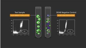
Prognostic significance of tumorigenic cells with mesenchymal features in pancreatic adenocarcinoma.
BACKGROUND: Specific populations of highly tumorigenic cells are thought to exist in many human tumors,including pancreatic adenocarcinoma. However,the clinical significance of these tumor-initiating (ie,cancer stem) cells remains unclear. Aldehyde dehydrogenase (ALDH) activity can identify tumor-initiating cells and normal stem cells from several human tissues. We examined the prognostic significance and functional features of ALDH expression in pancreatic adenocarcinoma. METHODS: ALDH expression was analyzed by immunohistochemistry in 269 primary surgical specimens of pancreatic adenocarcinoma and examined for association with clinical outcomes and in paired primary tumors and metastatic lesions from eight pancreatic cancer patients who had participated in a rapid autopsy program. The clonogenic growth potential of ALDH-positive pancreatic adenocarcinoma cells was assessed in vitro by a colony formation assay and by tumor growth in immunodeficient mice (10-14 mice per group). Mesenchymal features of ALDH-positive pancreatic tumor cells were examined by using quantitative reverse transcription-polymerase chain reaction and an in vitro cell invasion assay. Gene expression levels and the invasive potential of ADLH-positive pancreatic cancer cells relative to the bulk cell population were examined by reverse transcription-polymerase chain reaction and an in vitro invasion assays,respectively. All statistical tests were two-sided. RESULTS: ALDH-positive tumor cells were detected in 90 of the 269 primary surgical specimens,and their presence was associated with worse survival (median survival for patients with ALDH-positive vs ALDH-negative tumors: 14 vs 18 months,hazard ratio of death = 1.28,95% confidence interval = 1.02 to 1.68,P = .05). Six (75%) of the eight patients with matched primary and metastatic tumor samples had ALDH-negative primary tumors,and in four (67%) of these six patients,the matched metastatic lesions (located in liver and lung) contained ALDH-positive cells. ALDH-positive cells were approximately five- to 11-fold more clonogenic in vitro and in vivo compared with unsorted or ALHD-negative cells,expressed genes consistent with a mesenchymal state,and had in vitro migratory and invasive potentials that were threefold greater than those of unsorted cells. CONCLUSIONS: ALDH expression marks pancreatic cancer cells that have stem cell and mesenchymal features. The enhanced clonogenic growth and migratory properties of ALDH-positive pancreatic cancer cells suggest that they play a key role in the development of metastatic disease that negatively affects the overall survival of patients with pancreatic adenocarcinoma.
View Publication
产品号#:
01700
01705
01702
产品名:
ALDEFLUOR™ 试剂盒
ALDEFLUOR™ DEAB试剂, 1.5 mM, 1 mL
ALDEFLUOR™检测缓冲液
Maes C et al. (MAY 2006)
The Journal of clinical investigation 116 5 1230--42
Placental growth factor mediates mesenchymal cell development, cartilage turnover, and bone remodeling during fracture repair.
Current therapies for delayed- or nonunion bone fractures are still largely ineffective. Previous studies indicated that the VEGF homolog placental growth factor (PlGF) has a more significant role in disease than in health. Therefore we investigated the role of PlGF in a model of semi-stabilized bone fracture healing. Fracture repair in mice lacking PlGF was impaired and characterized by a massive accumulation of cartilage in the callus,reminiscent of delayed- or nonunion fractures. PlGF was required for the early recruitment of inflammatory cells and the vascularization of the fracture wound. Interestingly,however,PlGF also played a role in the subsequent stages of the repair process. Indeed in vivo and in vitro findings indicated that PlGF induced the proliferation and osteogenic differentiation of mesenchymal progenitors and stimulated cartilage turnover by particular MMPs. Later in the process,PlGF was required for the remodeling of the newly formed bone by stimulating osteoclast differentiation. As PlGF expression was increased throughout the process of bone repair and all the important cell types involved expressed its receptor VEGFR-1,the present data suggest that PlGF is required for mediating and coordinating the key aspects of fracture repair. Therefore PlGF may potentially offer therapeutic advantages for fracture repair.
View Publication
产品号#:
03534
03334
03434
03444
18753
18753RF
产品名:
MethoCult™ GF M3534
MethoCult™ M3334
MethoCult™ GF M3434
MethoCult™ GF M3434
Eirew P et al. (DEC 2008)
Nature medicine 14 12 1384--9
A method for quantifying normal human mammary epithelial stem cells with in vivo regenerative ability.
Previous studies have demonstrated that normal mouse mammary tissue contains a rare subset of mammary stem cells. We now describe a method for detecting an analogous subpopulation in normal human mammary tissue. Dissociated cells are suspended with fibroblasts in collagen gels,which are then implanted under the kidney capsule of hormone-treated immunodeficient mice. After 2-8 weeks,the gels contain bilayered mammary epithelial structures,including luminal and myoepithelial cells,their in vitro clonogenic progenitors and cells that produce similar structures in secondary transplants. The regenerated clonogenic progenitors provide an objective indicator of input mammary stem cell activity and allow the frequency and phenotype of these human mammary stem cells to be determined by limiting-dilution analysis. This new assay procedure sets the stage for investigations of mechanisms regulating normal human mammary stem cells (and possibly stem cells in other tissues) and their relationship to human cancer stem cell populations.
View Publication
产品号#:
05601
产品名:
EpiCult™-B 人培养基
Lindvall C et al. (NOV 2006)
The Journal of biological chemistry 281 46 35081--7
The Wnt signaling receptor Lrp5 is required for mammary ductal stem cell activity and Wnt1-induced tumorigenesis.
Canonical Wnt signaling has emerged as a critical regulatory pathway for stem cells. The association between ectopic activation of Wnt signaling and many different types of human cancer suggests that Wnt ligands can initiate tumor formation through altered regulation of stem cell populations. Here we have shown that mice deficient for the Wnt co-receptor Lrp5 are resistant to Wnt1-induced mammary tumors,which have been shown to be derived from the mammary stem/progenitor cell population. These mice exhibit a profound delay in tumorigenesis that is associated with reduced Wnt1-induced accumulation of mammary progenitor cells. In addition to the tumor resistance phenotype,loss of Lrp5 delays normal mammary development. The ductal trees of 5-week-old Lrp5-/- females have fewer terminal end buds,which are structures critical for juvenile ductal extension presumed to be rich in stem/progenitor cells. Consequently,the mature ductal tree is hypomorphic and does not completely fill the fat pad. Furthermore,Lrp5-/- ductal cells from mature females exhibit little to no stem cell activity in limiting dilution transplants. Finally,we have shown that Lrp5-/- embryos exhibit substantially impaired canonical Wnt signaling in the primitive stem cell compartment of the mammary placodes. These findings suggest that Lrp5-mediated canonical signaling is required for mammary ductal stem cell activity and for tumor development in response to oncogenic Wnt effectors.
View Publication

 EasySep™小鼠TIL(CD45)正选试剂盒


 沪公网安备31010102008431号