Lymphodepletion in the ApcMin/+ mouse model of intestinal tumorigenesis.
Germ line mutations in the Adenomatous polyposis coli tumor suppressor gene cause a hereditary form of intestinal tumorigenesis in both mice and man. Here we show that in Apc(Min/+) mice,which carry a heterozygous germ line mutation at codon 850 of Apc,there is progressive loss of immature and mature thymocytes from approximately 80 days of age with complete regression of the thymus by 120 days. In addition,Apc(Min/+) mice show parallel depletion of splenic natural killer (NK) cells,immature B cells,and B progenitor cells in bone marrow due to complete loss of interleukin 7 (IL-7)-dependent B-cell progenitors. Using bone marrow transplantation experiments into wild-type recipients,we have shown that the capacity of transplanted Apc(Min/+) bone marrow cells for T- and B-cell development appears normal. In contrast,although the Apc(Min/+) bone marrow microenvironment supported short-term reconstitution with wild-type bone marrow,Apc(Min/+) animals that received transplants subsequently underwent lymphodepletion. Fibroblast colony-forming unit (CFU-F) colony assays revealed a significant reduction in colony-forming mesenchymal progenitor cells in the bone marrow of Apc(Min/+) mice compared with wild-type animals prior to the onset of lymphodepletion. This suggests that an altered bone marrow microenvironment may account for the selective lymphocyte depletion observed in this model of familial adenomatous polyposis.
View Publication
产品号#:
03630
03434
03444
05501
05502
产品名:
MethoCult™ M3630
MethoCult™ GF M3434
MethoCult™ GF M3434
Tripp A et al. (NOV 2003)
Journal of virology 77 22 12152--64
Human T-cell leukemia virus type 1 tax oncoprotein suppression of multilineage hematopoiesis of CD34+ cells in vitro.
Human T-cell leukemia virus type 1 (HTLV-1) and HTLV-2 are highly related viruses that differ in disease manifestation. HTLV-1 is the etiologic agent of adult T-cell leukemia and lymphoma,an aggressive clonal malignancy of human CD4-bearing T lymphocytes. Infection with HTLV-2 has not been conclusively linked to lymphoproliferative disorders. We previously showed that human hematopoietic progenitor (CD34(+)) cells can be infected by HTLV-1 and that proviral sequences were maintained after differentiation of infected CD34(+) cells in vitro and in vivo. To investigate the role of the Tax oncoprotein of HTLV on hematopoiesis,bicistronic lentiviral vectors were constructed encoding the HTLV-1 or HTLV-2 tax genes (Tax1 and Tax2,respectively) and the green fluorescent protein marker gene. Human hematopoietic progenitor (CD34(+)) cells were infected with lentivirus vectors,and transduced cells were cultured in a semisolid medium permissive for the development of erythroid,myeloid,and primitive progenitor colonies. Tax1-transduced CD34(+) cells displayed a two- to fivefold reduction in the total number of hematopoietic clonogenic colonies that arose in vitro,in contrast to Tax2-transduced cells,which showed no perturbation of hematopoiesis. The ratio of colony types that developed from Tax1-transduced CD34(+) cells remained unaffected,suggesting that Tax1 inhibited the maturation of relatively early,uncommitted hematopoietic stem cells. Since previous reports have linked Tax1 expression with initiation of apoptosis,lentiviral vector-mediated transduction of Tax1 or Tax2 was investigated in CEM and Jurkat T-cell lines. Ectopic expression of either Tax1 or Tax2 failed to induce apoptosis in T-cell lines. These data demonstrate that Tax1 expression perturbs development and maturation of pluripotent hematopoietic progenitor cells,an activity that is not displayed by Tax2,and that the suppression of hematopoiesis is not attributable to induction of apoptosis. Since hematopoietic progenitor cells may serve as a latently infected reservoir for HTLV infection in vivo,the different abilities of HTLV-1 and -2 Tax to suppress hematopoiesis may play a role in the respective clinical outcomes after infection with HTLV-1 or -2.
View Publication
产品号#:
02690
产品名:
StemSpan™ CC100
Yang W-T and Zheng P-S (FEB 2014)
PloS one 9 2 e88827
Promoter hypermethylation of KLF4 inactivates its tumor suppressor function in cervical carcinogenesis.
OBJECTIVE The KLF4 gene has been shown to be inactivated in cervical carcinogenesis as a tumor suppressor. However,the mechanism of KLF4 silencing in cervical carcinomas has not yet been identified. DNA methylation plays a key role in stable suppression of gene expression. METHODS The methylation status of the KLF4 promoter CpG islands was analyzed by bisulfite sequencing (BSQ) in tissues of normal cervix and cervical cancer. KLF4 gene expression was detected by RT-PCR,immunohistochemistry and western blot. KLF4 promoter methylation in cervical cancer cell line was determined by BSQ and methylation-specific polymerase chain reaction (MS-PCR). Cell proliferation ability was detected by cell growth curve and MTT assay. RESULTS The methylated allele was found in 41.90% of 24 cervical cancer tissues but only in 11.11% of 11 normal cervix tissues (Ptextless0.005). KLF4 mRNA levels were significantly reduced in cervical cancer tissues compared with normal cervix tissues (Ptextless0.01) and KLF4 mRNA expression showed a significant negative correlation with the promoter hypermethylation (r = -0.486,P = 0.003). Cervical cancer cell lines also showed a significant negative correlation between KLF4 expression and hypermethylation. After treatment with the demethylating agent 5-Azacytidine (5-Aza),the expression of KLF4 in the cervical cancer cell lines at both mRNA and protein levels was drastically increased,the cell proliferation ability was inhibited and the chemosensitivity for cisplatin was significantly increased. CONCLUSION KLF4 gene is inactivated by methylation-induced silencing mechanisms in a large subset of cervical carcinomas and KLF4 promoter hypermethylation inactivates the gene's function as a tumor suppressor in cervical carcinogenesis.
View Publication
Schmidt K et al. (MAR 2009)
Journal of leukocyte biology 85 3 563--73
Histone deacetylase inhibition improves differentiation of dendritic cells from leukemic blasts of patients with TEL/AML1-positive acute lymphoblastic leukemia.
Histone deacetylase inhibitors (HdI) could potentially improve the differentiation of leukemic dendritic cells (DC). Therefore,bone marrow samples from 100 children with acute lymphoblastic leukemia (ALL) were cultured in the cytokines TNF-alpha,GM-CSF,c-kit ligand,and fetal liver tyrosine kinase 3 ligand,with or without IL-3 and -4 and after administration of HdI valproic acid (VAL),suberoylanilide hydroxamic acid (SAHA),isobutyramid,or trichostatin A. Among the tested samples,25 were positive for the chromosomal translocation t(12;21),encoding the fusion gene translocation ETS-like leukemia/acute myeloid leukemia 1 (TEL/AML1). SAHA increased CD83 expression of TEL/AML1-positive blasts in conditions without ILs,and SAHA and VAL increased the number of CD86(+)80(-) cells in the presence of ILs. VAL and isobutyramid supported the allostimulatory capacities of TEL/AML1-positive,leukemic DC; VAL and SAHA reduced those of TEL/AML1-negative DC. Cytotoxic T cells sensitized with leukemic DC produced more IFN-gamma and TNF-alpha upon presentation of the TEL/AML1 peptide. They also induced the cytotoxic lysis of nondifferentiated blasts,which was enhanced when TEL/AML1-positive DC had developed after addition of VAL or SAHA. Therefore,the use of HdI in the differentiation of leukemic DC from patients with TEL/AML1-positive ALL is recommended.
View Publication
Kordon EC and Smith GH (MAY 1998)
Development (Cambridge,England) 125 10 1921--30
An entire functional mammary gland may comprise the progeny from a single cell.
Any epithelial portion of a normal mouse mammary gland can reproduce an entire functional gland when transplanted into an epithelium-free mammary fat pad. Mouse mammary hyperplasias and tumors are clonal dominant populations and probably represent the progeny of a single transformed cell. Our study provides evidence that single multipotent stem cells positioned throughout the mature fully developed mammary gland have the capacity to produce sufficient differentiated progeny to recapitulate an entire functional gland. Our evidence also demonstrates that these stem cells are self-renewing and are found with undiminished capacities in the newly regenerated gland. We have taken advantage of an experimental model where mouse mammary tumor virus infects mammary epithelial cells and inserts a deoxyribonucleic acid copy(ies) of its genome during replication. The insertions occur randomly within the somatic genome. CzechII mice have no endogenous nucleic acid sequence homology with mouse mammary tumor virus; therefore all viral insertions may be detected by Southern analysis provided a sufficient number of cells contain a specific insertional event. Transplantation of random fragments of infected CzechII mammary gland produced clonal-dominant epithelial populations in epithelium-free mammary fat pads. Serial transplantation of pieces of the clonally derived outgrowths produced second generation glands possessing the same viral insertion sites providing evidence for self-renewal of the original stem cell. Limiting dilution studies with cell cultures derived from third generation clonal outgrowths demonstrated that three multipotent but distinct mammary epithelial progenitors were present in clonally derived mammary epithelial populations. Estimation of the potential number of multipotent epithelial cells that may be evolved from an individual mammary-specific stem cell by self-renewal is in the order of 10(12)-10(13). Therefore,one stem cell might easily account for the renewal of mammary epithelium over several transplant generations.
View Publication
产品号#:
01700
01705
05601
05610
05620
01702
产品名:
ALDEFLUOR™ 试剂盒
ALDEFLUOR™ DEAB试剂, 1.5 mM, 1 mL
EpiCult™-B 人培养基
EpiCult™-B 小鼠培养基
MammoCult™ 人源培养基套装
ALDEFLUOR™检测缓冲液
Risueñ et al. (JUN 2011)
Blood 117 26 7112--20
Identification of T-lymphocytic leukemia-initiating stem cells residing in a small subset of patients with acute myeloid leukemic disease.
Xenotransplantation of acute myeloid leukemia (AML) into immunodeficient mice has been critical for understanding leukemogenesis in vivo and defining self-renewing leukemia-initiating cell subfractions (LICs). Although AML-engraftment capacity is considered an inherent property of LICs,substrains of NOD/SCID mice that possess additional deletions such as the IL2Rγc(null) (NSG) have been described as a more sensitive recipient to assay human LIC function. Using 23 AML-patient samples,39% demonstrated no detectable engraftment in NOD/SCID and were categorized as AMLs devoid of LICs. However,33% of AML patients lacking AML-LICs were capable of engrafting NSG recipients,but produced a monoclonal T-cell proliferative disorder similar to T-ALL. These grafts demonstrated self-renewal capacity as measured by in vivo serial passage and were restricted to CD34-positive fraction,and were defined as LICs. Molecular analysis for translocations in MLL genes indicated that these AML patient-derived LICs all expressed the MLL-AFX1 fusion product. Our results reveal that the in vivo human versus xenograft host microenvironment dictates the developmental capacity of human LICs residing in a small subset of patients diagnosed with AML harboring MLL mutations. These findings have implications both for the basic biology of CSC function,and for the use of in vivo models of the leukemogenic process in preclinical or diagnostic studies.
View Publication


 EasySep™小鼠TIL(CD45)正选试剂盒







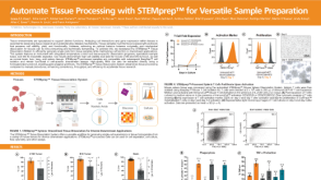 科学海报Automate Tissue Processing with STEMprep™ for Versatile Sample Preparation

 沪公网安备31010102008431号